Name this structure
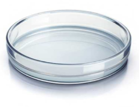
Petri dish
Agar plate
The Correct Answer is A
A Petri dish is a shallow, cylindrical, lidded container commonly used in laboratories for culturing microorganisms such as bacteria, fungi, and cells. It is typically made of clear glass or disposable sterile plastic, which allows easy observation of microbial growth. The dish consists of two parts: a flat bottom portion that holds the growth medium and a slightly larger lid that covers it to protect the culture from contamination while still allowing gas exchange. In microbiology, the bottom of the dish is usually filled with a nutrient medium such as agar that supports microbial growth. Samples are inoculated onto the agar surface and incubated under controlled temperature conditions. As microorganisms multiply
Nursing Test Bank
Naxlex Comprehensive Predictor Exams
Related Questions
Correct Answer is A
Explanation
The fine adjustment knob on a microscope is used to sharpen or clarify the image after the initial focus has been achieved with the coarse adjustment knob. The coarse adjustment knob moves the stage or objective lens quickly over a larger distance to bring the specimen roughly into focus, which is especially useful with low-power objectives. Once the image is visible, the fine adjustment knob allows for precise, gradual adjustments to enhance the clarity and detail of the specimen, particularly under high-power or oil immersion objectives. This ensures accurate visualization of cellular structures without damaging the slide or lens.
Correct Answer is B
Explanation
A. Their outer membrane traps the stain: Gram-positive bacteria do not have an outer membrane; this structure is characteristic of Gram-negative bacteria. Therefore, the outer membrane cannot be responsible for retaining crystal violet in Gram-positive cells.
B. Their thick peptidoglycan layer holds the crystal violet-iodine complex: Gram-positive bacteria have a thick, multilayered peptidoglycan cell wall that forms a dense network. During Gram staining, the crystal violet combines with iodine to form an insoluble complex that gets trapped within this thick peptidoglycan matrix. Even after alcohol or acetone decolorization, the complex remains, causing the cells to appear purple under a microscope.
C. They absorb the safranin more strongly: Safranin is a counterstain used to color Gram-negative bacteria red after decolorization. Gram-positive bacteria do not absorb safranin strongly because the retained crystal violet-iodine complex masks it. Therefore, safranin absorption is not responsible for the purple color of Gram-positive cells.
D. They do not undergo decolorization: While Gram-positive bacteria resist decolorization, this is not because they fail to undergo the process; it is due to the structural ability of their thick peptidoglycan to trap the crystal violet-iodine complex. The resistance is a result of the cell wall structure, not a lack of exposure to the decolorizing agent.
Whether you are a student looking to ace your exams or a practicing nurse seeking to enhance your expertise , our nursing education contents will empower you with the confidence and competence to make a difference in the lives of patients and become a respected leader in the healthcare field.
Visit Naxlex, invest in your future and unlock endless possibilities with our unparalleled nursing education contents today