
Please identify the following rhythm: 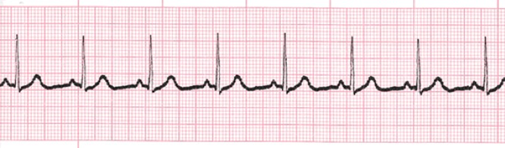
Asystole
Atrial Flutter
Normal Sinus Rhythm
Sinus Bradycardia
The Correct Answer is C
Answer and explanation
The correct answer is C. Normal Sinus Rhythm.
Choice A rationale:
Asystole is the absence of all electrical activity in the heart, as evidenced by a flat line on the electrocardiogram (ECG). It is a medical emergency that requires immediate cardiopulmonary resuscitation (CPR) and defibrillation.
Key features of asystole on ECG:
No discernible P waves, QRS complexes, or T waves.
A completely flat or nearly flat line on the ECG tracing.
Choice B rationale:
Atrial flutter is a rapid heart rhythm that arises from abnormal electrical activity in the atria. It is characterized by a sawtooth pattern on the ECG, with atrial rates typically between 250 and 350 beats per minute.
Key features of atrial flutter on ECG:
Absence of distinct P waves, instead replaced by flutter waves (sawtooth pattern).
Regular, rapid atrial rate (typically 250-350 bpm).
QRS complexes may be normal or slightly irregular in appearance.
Choice C rationale:
Normal sinus rhythm is the natural, healthy rhythm of the heart. It originates in the sinoatrial (SA) node, the heart's natural pacemaker, and is characterized by a regular rate of 60-100 beats per minute, with consistent P waves, QRS complexes, and T waves on the ECG.
Key features of normal sinus rhythm on ECG:
Presence of distinct P waves, QRS complexes, and T waves.
Regular rhythm with a rate of 60-100 beats per minute.
PR interval (the time between the P wave and QRS complex) is 0.12-0.20 seconds.
QRS duration (the time it takes for the ventricles to depolarize) is less than 0.12 seconds.
Choice D rationale:
Sinus bradycardia is a slow heart rhythm, with a rate below 60 beats per minute. It is often a normal finding in healthy individuals, especially athletes or during sleep. However, it can also be a sign of underlying medical conditions.
Key features of sinus bradycardia on ECG:
Presence of distinct P waves, QRS complexes, and T waves.
Regular rhythm with a rate less than 60 beats per minute.
PR interval and QRS duration are typically normal.
Nursing Test Bank
Naxlex Comprehensive Predictor Exams
Related Questions
Correct Answer is ["A","C","D"]
Explanation
Rationale for Choice A:
Monitoring the insertion site for hematoma and bleeding is crucial following cardiac angiogram with stent placement. Here's a detailed explanation:
Vascular Injury: The procedure involves accessing a major artery, usually the femoral artery in the groin. This creates a risk of bleeding or hematoma formation at the puncture site.
Anticoagulation: Patients often receive anticoagulant medications during and after the procedure to prevent blood clots from forming around the stent. These medications can increase the risk of bleeding.
Early Detection and Intervention: Prompt identification of bleeding or hematoma formation is essential to prevent complications such as hypotension, shock, or compartment syndrome.
Assessment: Regular assessment of the insertion site includes:
Visual inspection for swelling, bruising, or active bleeding.
Palpation for tenderness, firmness, or expanding hematoma.
Monitoring vital signs for tachycardia, hypotension, or signs of shock.
Interventions: If bleeding or hematoma is detected:
Apply direct pressure to the site.
Notify the healthcare provider immediately.
Implement additional measures as prescribed, such as reversal of anticoagulation or surgical intervention.
Rationale for Choice B:
Maintaining strict bedrest after cardiac angiogram with stent placement is no longer routinely recommended. Here's why:
Early Ambulation Benefits: Research has shown that early ambulation (usually within 6-8 hours) is associated with several benefits, including:
Reduced risk of deep vein thrombosis (DVT)
Improved circulation
Quicker recovery
Shorter hospital stays
Individualized Assessment: The decision regarding ambulation timing should be individualized based on patient factors such as:
Bleeding risk
Procedural complications
Overall health status
Comfort level
Balanced Approach: While early ambulation is encouraged, it's important to balance activity with rest to promote healing and prevent complications.
Rationale for Choice C:
Monitoring for dysrhythmias is essential following cardiac angiogram with stent placement due to several reasons:
Cardiac Irritation: The procedure involves manipulation of catheters and devices within the heart, which can potentially irritate the heart's electrical system.
Electrolyte Imbalances: Contrast dye used during the procedure can cause electrolyte imbalances, which can also trigger dysrhythmias.
Underlying Heart Disease: Patients undergoing angioplasty often have underlying heart disease, which already predisposes them to rhythm disturbances.
Early Detection and Intervention: Prompt identification of dysrhythmias allows for timely interventions to prevent hemodynamic instability, heart failure, or cardiac arrest.
Continuous Monitoring: Continuous ECG monitoring is typically used to detect dysrhythmias, and patients are closely observed for symptoms such as palpitations, lightheadedness, or chest pain.
Rationale for Choice D:
Assessing heart sounds is a crucial nursing intervention following cardiac angiogram with stent placement. Here's why:
Stent Placement Effects: The placement of a stent can alter blood flow patterns within the coronary arteries, which may be audible as changes in heart sounds.
Complication Detection: Heart sounds can provide valuable clues about potential complications such as:
Pericardial effusion (fluid around the heart)
Myocardial infarction (heart attack)
Valvular dysfunction
Baseline Assessment: It's important to establish a baseline assessment of heart sounds prior to the procedure to compare with post-procedure findings.
Regular Auscultation: Regular auscultation of heart sounds should be performed to monitor for changes and promptly report any concerns to the healthcare provider.
Correct Answer is B
Explanation
Choice B rationale:
LDL (low-density lipoprotein) cholesterol is often referred to as "bad" cholesterol. It contributes to the buildup of plaque in the arteries, which narrows the arteries and can lead to coronary artery disease (CAD). CAD is a major risk factor for heart attack and stroke.
HDL (high-density lipoprotein) cholesterol is often referred to as "good" cholesterol. It helps to remove LDL cholesterol from the arteries and transport it back to the liver, where it can be broken down and removed from the body. Therefore, a high HDL level is protective against heart disease.
An elevated LDL level and a lowered HDL level are both associated with an increased risk of CAD. This is because the elevated LDL level contributes to plaque buildup, while the lowered HDL level means that there is less "good" cholesterol available to remove the "bad" cholesterol from the arteries.
Therefore, the statement "Increased LDL and decreased HDL increase my risk of coronary artery disease" indicates an understanding of the significance of these cholesterol levels.
Choice A rationale:
This statement is incorrect. An increased LDL level will actually increase the amount of cholesterol deposited on the artery walls.
Choice C rationale:
This statement is also incorrect. A decreased HDL level will decrease the amount of cholesterol moved away from the artery walls.
Choice D rationale:
This statement is incorrect. Increased LDL has the potential to increase, not decrease, the risk of heart disease.
Whether you are a student looking to ace your exams or a practicing nurse seeking to enhance your expertise , our nursing education contents will empower you with the confidence and competence to make a difference in the lives of patients and become a respected leader in the healthcare field.
Visit Naxlex, invest in your future and unlock endless possibilities with our unparalleled nursing education contents today