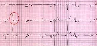
Premature ventricular contractions (PVCs) occur while the nurse is suctioning a patient's endotracheal tube. Which action by the nurse is best?
Stop and ventilate the patient with 100% oxygen.
Check the patient's potassium level
Give prescribed PRN antidysrhythmic medications.
Decrease the suction pressure to 80 mm Hg.
The Correct Answer is A
Premature ventricular contractions (PVCs) are abnormal heart rhythms originating from the ventricles. They can be triggered by various factors, including irritation or stimulation of the airway during suctioning.
In this situation, the priority is to ensure adequate oxygenation and ventilation for the patient. Stopping the suctioning procedure and providing ventilatory support with 100% oxygen helps maintain oxygen levels and minimizes further cardiac dysrhythmias.
B. Check the patient's potassium level in (option B) is incorrect because While electrolyte imbalances, including low potassium levels (hypokalemia), can contribute to cardiac dysrhythmias, checking the potassium level is not the immediate priority when PVCs occur during suctioning.
C. Give prescribed PRN antidysrhythmic medications in (option C) is incorrect because: Administering antidysrhythmic medications without further assessment or evaluation may not be appropriate in this situation.
D. Decrease the suction pressure to 80 mm Hg in (option D) is incorrect because: While adjusting suction pressure may help prevent further irritation, it is not the initial priority when PVCs are present during suctioning.
E. Documenting the dysrhythmia in the patient's chart in (option E) is incorrect because: Documentation is important but should not be the initial action when a patient experiences PVCs during suctioning. Patient safety and immediate intervention take precedence.
Therefore, when PVCs occur during suctioning, the nurse should stop the procedure, provide ventilatory support with 100% oxygen, and assess the patient's response to intervention.
Nursing Test Bank
Naxlex Comprehensive Predictor Exams
Related Questions
Correct Answer is C
Explanation
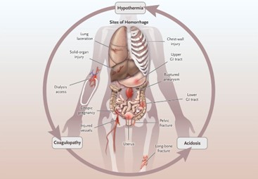
Hemorrhagic shock is characterized by severe blood loss, leading to inadequate tissue perfusion and hypovolemia. The primary goal in the initial management of hemorrhagic shock is to restore intravascular volume and improve tissue perfusion. Administering intravenous fluids, such as normal saline solution, is a critical intervention to address hypovolemia and improve blood pressure.
A. Give Plasmanate 1 unit now in (option A) is incorrect because: Plasmanate is a plasma-derived product used to replace coagulation factors. While it may be necessary to address coagulation abnormalities, administering intravenous fluids to restore volume takes priority over specific blood products.
B. Prepare for endotracheal intubation in (option B) is incorrect because Endotracheal intubation may be required in cases of impending respiratory failure or compromised airway, but it should not be the first action in addressing hypovolemic shock.
D. Type and crossmatch for 4 units of packed red blood cells (PRBCs) in (option D) is incorrect because transferring packed red blood cells is an important intervention to address blood loss and improve oxygen-carrying capacity. However, before administering blood products, it is crucial to stabilize the patient's hemodynamics through fluid resuscitation.
Therefore, in a patient with hemorrhagic shock, the nurse's first priority among the given options is to give normal saline solution of 250 mL/hr to restore intravascular volume and improve tissue perfusion.
Correct Answer is B
Explanation
Positioning the transducer level with the phlebostatic axis is a crucial step in accurate hemodynamic monitoring. The phlebostatic axis is an imaginary reference point located at the fourth intercostal space, mid-anterior/posterior chest. Placing the transducer at this level ensures that the pressure measurements obtained are reflective of the patient's true hemodynamic status.
A. Positioning the limb with the catheter insertion site at the level of the transducer in (option A) is incorrect because: While it is important to position the limb appropriately to avoid kinks or occlusions in the catheter tubing, this is not directly related to the accurate measurement of hemodynamic parameters.
C. Ensuring that the patient is lying with the head of the bed flat for all readings in (option C) is incorrect because The position of the patient's head does not directly impact the accuracy of hemodynamic monitoring unless it specifically relates to changes in preload or intracranial pressure monitoring.
D. Balancing and calibrating the hemodynamic monitoring equipment every hour in (option D) is incorrect because: While it is important to ensure that the monitoring equipment is calibrated and functioning properly, doing so every hour may not be necessary. Calibration frequency may vary based on institutional policies and patient stability.
Therefore, the correct action that demonstrates effective teaching about hemodynamic monitoring is positioning the transducer level with the phlebostatic axis.
Whether you are a student looking to ace your exams or a practicing nurse seeking to enhance your expertise , our nursing education contents will empower you with the confidence and competence to make a difference in the lives of patients and become a respected leader in the healthcare field.
Visit Naxlex, invest in your future and unlock endless possibilities with our unparalleled nursing education contents today