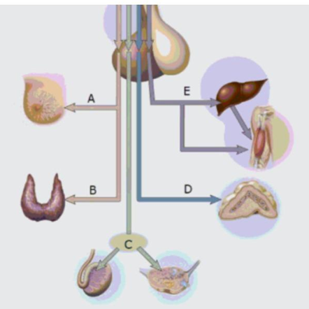
What is the name of the hormone at E?
Growth hormone
Prolactin
Thyroid stimulating hormone
Gonadotropins
ACTH
The Correct Answer is A
A. Hormone at E - Growth Hormone (GH): The diagram indicates that hormone E is released from the anterior pituitary and targets the liver and skeletal muscles. In the liver, it stimulates the production of insulin-like growth factors (IGFs) to promote systemic growth. Its action on skeletal muscle facilitates protein synthesis and tissue hypertrophy.
B. Hormone A - Prolactin (PRL): The diagram depicts hormone A traveling from the adenohypophysis to the mammary glands. This protein hormone is essential for initiating and maintaining milk production following parturition. Its secretion is regulated by hypothalamic dopamine, which serves as a primary prolactin-inhibiting factor.
C. Hormone B - Thyroid-stimulating Hormone (TSH): The pathway labeled B shows a tropic hormone targeting the thyroid gland. TSH stimulates the follicular cells of the thyroid to synthesize and release thyroxine and triiodothyronine. This hormone is a critical regulator of systemic basal metabolic rate and cellular heat production.
D. Hormone C - Gonadotropins (FSH and LH): Label C represents the gonadotropins, specifically follicle-stimulating hormone and luteinizing hormone, which target the testes and ovaries. These hormones regulate gametogenesis and the secretion of sex steroids like testosterone and estrogen. They are essential for the maintenance of reproductive cycles and secondary sexual characteristics.
E. Hormone at D - Adrenocorticotropic Hormone (ACTH): The diagram shows hormone D being secreted from the anterior pituitary and traveling specifically to the adrenal gland. More specifically, it targets the adrenal cortex to regulate the production of steroid hormones. It is a critical component of the hypothalamic-pituitary-adrenal (HPA) axis.
Nursing Test Bank
Naxlex Comprehensive Predictor Exams
Related Questions
Correct Answer is C
Explanation
A. Carbaminohemoglobin: Approximately 23% of carbon dioxide is transported by binding to the globin amino groups of the hemoglobin molecule. While this is a significant transport mechanism, it is secondary to the ionic pathway in the plasma. It primarily occurs after oxygen is unloaded at the systemic tissues.
B. Carboxyhemoglobin: This term describes the stable complex formed when carbon monoxide, not carbon dioxide, binds to the heme iron of hemoglobin. This condition is pathological and interferes with oxygen transport. It is not a normal physiological mechanism for the excretion of metabolic carbon dioxide.
C. bicarbonate ions: About 70% of metabolic carbon dioxide is converted into bicarbonate ions within erythrocytes by the enzyme carbonic anhydrase. These ions then diffuse into the plasma for transport to the lungs. This represents the most voluminous and efficient method for carrying carbon dioxide in blood.
D. dissolved CO2 gas: Only about 7% to 10% of carbon dioxide is transported physically dissolved in the blood plasma. Due to the limited solubility of gases in aqueous solutions, this method cannot accommodate the high metabolic output of tissues. Most of the gas must be chemically converted to be carried.
E. bisphosphocarbonate: This molecule is not a recognized physiological transporter of carbon dioxide in human hematology. It may be confused with 2,3-bisphosphoglycerate, which regulates hemoglobin's oxygen affinity. It plays no role in the chemical buffering or transport of carbon dioxide within the circulatory system.
Correct Answer is B
Explanation
A. chief cells; carbonic anhydrase (CAH); parietal cells: Chief cells correctly synthesize the zymogen pepsinogen, but carbonic anhydrase is an enzyme, not a direct activator. CAH facilitates the formation of protons within cells but does not catalyze extracellular protein cleavage. Pepsinogen requires a low pH environment for activation.
B. chief cells; hydrochloric acid (HCl); parietal cells: Gastric chief cells secrete inactive pepsinogen into the stomach lumen. Hydrochloric acid, produced by parietal cells via proton pumps, lowers the luminal pH to approximately 2. This acidic environment triggers the autocatalytic conversion of pepsinogen into the active protease pepsin.
C. parietal cells; hydrochloric acid (HCl): chief cells: This selection incorrectly reverses the cellular origins of the enzyme and the acid. Parietal cells are responsible for secreting hydrochloric acid and intrinsic factor, not the zymogen pepsinogen. Chief cells provide the protein substrate but do not produce the acid required.
D. parietal cells; carbonic anhydrase (CAH); chief cells: Carbonic anhydrase is an intracellular enzyme that provides the hydrogen ions for acid production. It is not the molecule that directly interacts with pepsinogen in the gastric lumen. Furthermore, parietal cells do not produce the pepsinogen zymogen required for this reaction.
E. enteroendocrine cells; carbonic anhydrase (CAH); parietal cells: Enteroendocrine cells, specifically G cells, secrete hormones like gastrin into the bloodstream rather than digestive zymogens. Carbonic anhydrase remains an intracellular catalyst for ion formation. This combination fails to describe the luminal activation of proteases necessary for protein degradation.
Whether you are a student looking to ace your exams or a practicing nurse seeking to enhance your expertise , our nursing education contents will empower you with the confidence and competence to make a difference in the lives of patients and become a respected leader in the healthcare field.
Visit Naxlex, invest in your future and unlock endless possibilities with our unparalleled nursing education contents today