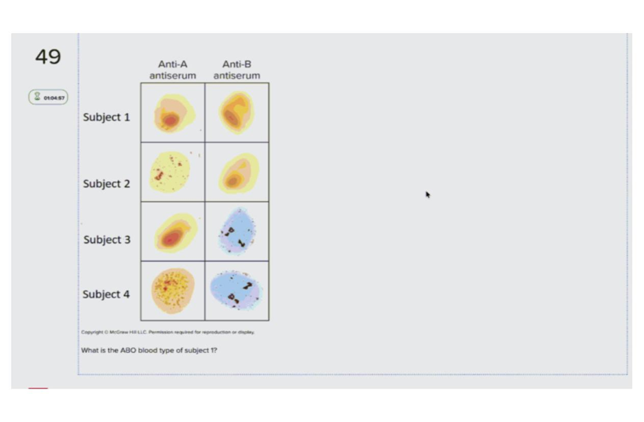
What is the ABO blood type of subject 1?
Correct answer: Blood group 0
Subject 1
Subject 2
Subject 3
Subject 4
The Correct Answer is A
A. Subject 1: Because Subject 1 lacks both A and B surface antigens, the individual is classified as having blood type O. In the ABO system, type O is characterized by the absence of these specific glycoproteins. This phenotype results in the presence of both anti-A and anti-B antibodies in the plasma.
B. Subject 2: The sample demonstrates visible agglutination in the presence of Anti-A antiserum while remaining smooth in Anti-B antiserum. This indicates the presence of A antigens and the absence of B antigens on the erythrocyte membranes. Consequently, the ABO blood group for this individual is Type A.
C. Subject 3: There is a clear lack of agglutination in the Anti-A antiserum, but distinct clumping is present in the Anti-B antiserum. The reaction confirms that the red blood cells possess B antigens but lack A antigens. This specific reactivity pattern identifies the individual as having Type B blood.
D. Subject 4: Agglutination is prominently visible in both the Anti-A and Anti-B antiserum wells for this individual. This positive reaction in both tests proves the simultaneous presence of both A and B surface antigens. Therefore, Subject 4 is classified as having blood type AB.
Nursing Test Bank
Naxlex Comprehensive Predictor Exams
Related Questions
Correct Answer is C
Explanation
A. visceral layers of the serous pericardium; parietal layers of the serous pericardium: These terms describe the thin, double-layered membrane that surrounds and protects the heart, forming the pericardial cavity. They are not chambers of the heart but rather protective coverings that facilitate frictionless movement. This choice incorrectly identifies serous membranes as anatomical internal heart cavities.
B. ventricles; atria: This selection inverts the correct anatomical arrangement of the human heart. The ventricles are the thick-walled, inferior pumping chambers that propel blood out into the pulmonary and systemic circuits. The atria are located superiorly to the ventricles and serve as the receiving chambers for returning blood.
C. atria; ventricles: The heart is divided into four chambers, with the two atria serving as the superior receiving chambers and the two ventricles as the inferior pumping chambers. The atria are separated from the ventricles by atrioventricular valves to ensure unidirectional blood flow. This accurately describes the vertical spatial relationship of the heart's internal anatomy.
D. left ventricles; right ventricles: These are the two inferior pumping chambers of the heart, located side-by-side rather than in a superior-inferior arrangement. While they differ in wall thickness and pressure output, both are situated below the level of the atria. They are separated by the thick interventricular septum.
E. left atria; right atria: These represent the two superior receiving chambers of the heart, divided by the interatrial septum. Like the ventricles, they are positioned horizontally relative to one another rather than vertically. They are both located superior to their respective ventricles within the thoracic cavity.
Correct Answer is B
Explanation
A. Tongue: This muscular organ facilitates mechanical digestion and bolus formation within the oral cavity. It contains gustatory receptors and serous glands but lacks hepatocytes for biochemical synthesis. It does not participate in the production or secretion of biliary salts or pigments.
B. Liver: Hepatocytes synthesize bile acids from cholesterol to facilitate the emulsification of dietary lipids. This accessory organ secretes the fluid into the biliary tree for eventual transport to the duodenum. It is the primary site for the biochemical production of bile.
C. Pancreas: This dual-function gland secretes alkaline juice containing digestive enzymes and bicarbonate into the small intestine. Its exocrine component focuses on proteases, lipases, and amylases rather than bile. It regulates blood glucose via endocrine secretions but does not produce biliary fluids.
D. Salivary glands: These exocrine glands produce saliva containing ptyalin and lingual lipase for initial chemical digestion. They maintain oral hygiene and lubricate the food bolus for deglutition. They lack the specialized metabolic machinery required to synthesize bile acids or bilirubin.
E. Gallbladder: This hollow organ functions exclusively as a reservoir for the concentration and storage of bile. It undergoes cholecystokinin-induced contraction to release bile into the common bile duct. While it manages bile distribution, it possesses no secretory tissue for bile synthesis.
Whether you are a student looking to ace your exams or a practicing nurse seeking to enhance your expertise , our nursing education contents will empower you with the confidence and competence to make a difference in the lives of patients and become a respected leader in the healthcare field.
Visit Naxlex, invest in your future and unlock endless possibilities with our unparalleled nursing education contents today