Which structure is circled in the image below?
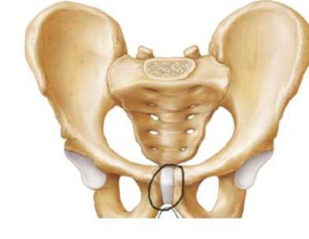
Pubic symphysis
Iliac crest
Ischial tuberosity
Sacroiliac joint
The Correct Answer is A
The marked structure is the pubic symphysis, a midline fibrocartilaginous joint located at the anterior aspect of the pelvis where the left and right pubic bones meet. It is composed of hyaline cartilage-covered pubic surfaces joined by an interpubic disc of fibrocartilage. This joint is classified as an amphiarthrosis, allowing only limited movement while providing strong stability to the anterior pelvic ring. It plays an important role in weight distribution during standing and walking and undergoes slight widening during childbirth due to hormonal influence (relaxin).
A. Pubic symphysis: The pubic symphysis is a cartilaginous joint located at the anterior midline of the pelvis, formed by the articulation of the left and right pubic bones. It contains a fibrocartilaginous disc that allows minimal movement, contributing to pelvic stability and shock absorption. It is particularly important in weight transfer between the lower limbs and axial skeleton. Since the circled structure is centrally located at the anterior pelvis, it corresponds to the pubic symphysis.
B. Iliac crest: The iliac crest is the superior curved border of the ilium, forming the prominent “hip bone” ridge that can be palpated on the lateral waist. It serves as an attachment site for abdominal muscles and fascia. Unlike the pubic symphysis, it is a broad lateral bony ridge rather than a midline joint structure.
C. Ischial tuberosity: The ischial tuberosity is a roughened, weight-bearing prominence on the inferior aspect of the ischium. It serves as the attachment site for the hamstring muscles and bears body weight during sitting. Compared to the pubic symphysis, it is posterior and inferior rather than anterior and midline.
D. Sacroiliac joint: The sacroiliac joint is the articulation between the sacrum and the ilium located posteriorly in the pelvis. It is a strong synovial joint that transfers weight from the axial skeleton to the lower limbs. Unlike the pubic symphysis, it is posterior and lateral rather than central at the anterior midline.
Nursing Test Bank
Naxlex Comprehensive Predictor Exams
Related Questions
Correct Answer is D
Explanation
The anterior cruciate ligament (ACL) is one of the major stabilizing ligaments of the knee joint. It is located within the joint capsule and extends from the anterior intercondylar area of the tibia to the posterior aspect of the lateral femoral condyle. The ACL prevents excessive anterior movement of the tibia relative to the femur and helps maintain rotational stability during activities such as running, jumping, and pivoting. ACL injuries are particularly common in athletes participating in sports that involve sudden changes in direction.
A. Shoulder: The shoulder joint is a ball-and-socket joint formed by the articulation of the humerus with the glenoid cavity of the scapula. Stability is provided by structures such as the rotator cuff muscles, glenohumeral ligaments, and labrum. The shoulder does not contain an anterior cruciate ligament. Therefore, an ACL injury cannot occur within the shoulder joint.
B. Hip: The hip is a weight-bearing ball-and-socket joint formed by the head of the femur and the acetabulum of the pelvis. It is stabilized by strong ligaments including the iliofemoral, pubofemoral, and ischiofemoral ligaments. These ligaments provide substantial support during standing and walking. The anterior cruciate ligament is not a component of the hip joint anatomy.
C. Ankle: The ankle joint is formed primarily by the tibia, fibula, and talus and is stabilized by ligaments such as the deltoid ligament medially and the anterior talofibular ligament laterally. Ankle sprains commonly involve these structures rather than cruciate ligaments. Since the ACL is not found in the ankle, this option is incorrect.
D. Knee: the anterior cruciate ligament is one of the four major ligaments of the knee joint, along with the posterior cruciate ligament, medial collateral ligament, and lateral collateral ligament. The ACL functions to prevent anterior displacement of the tibia and contributes significantly to rotational stability. Partial or complete ACL tears commonly occur during sports involving sudden deceleration, pivoting, or landing from a jump. Injury to this ligament specifically indicates damage to the knee joint.
Correct Answer is A
Explanation
The human body is organized into several major anatomical cavities that house and protect delicate internal organs. These cavities are lined by specific membranes and are categorized based on their location within the body's structural framework. The cranial cavity is a specialized dorsal space that acts as the protective bony container for the central nervous system, ensuring the brain remains shielded from external trauma.
A. The cranial cavity, also known as the endocranium, is the space enclosed by the bones of the skull. It directly houses the brain, the meninges, and the cerebrospinal fluid that cushions the brain. This cavity is continuous with the vertebral canal, forming the dorsal body cavity, which is the primary site of protection for the central nervous system.
B. The thoracic cavity is located in the upper trunk of the body, protected by the rib cage and the sternum. It houses vital organs such as the heart, lungs, esophagus, and trachea. It is separated from the abdominal cavity below by the diaphragm and is completely separate from the cranial cavity shown in the diagram.
C. The abdominal cavity occupies the upper portion of the abdominopelvic cavity. It contains the majority of the digestive organs, including the stomach, liver, intestines, and kidneys. Because it is located in the trunk and holds visceral organs associated with digestion and metabolism, it bears no anatomical relationship to the cranial cavity.
D. The pelvic cavity is the lowermost portion of the abdominopelvic cavity, located within the bony pelvis. It contains the urinary bladder, reproductive organs, and the terminal portions of the gastrointestinal tract. Like the abdominal cavity, it is positioned far from the head and is not involved in housing the brain.
Whether you are a student looking to ace your exams or a practicing nurse seeking to enhance your expertise , our nursing education contents will empower you with the confidence and competence to make a difference in the lives of patients and become a respected leader in the healthcare field.
Visit Naxlex, invest in your future and unlock endless possibilities with our unparalleled nursing education contents today