What is the classification of the highlighted bones?
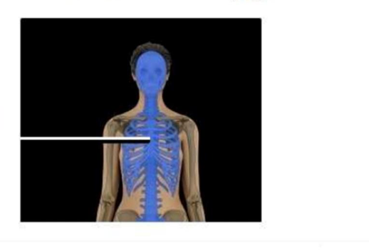
Appendicular skeleton
Axial skeleton
Irregular skeleton
Long bone group
The Correct Answer is B
The highlighted bones belong to the axial skeleton, which forms the central framework of the body. The axial skeleton includes the skull, vertebral column, and thoracic cage (ribs and sternum). Its primary function is to support and protect vital organs such as the brain, spinal cord, heart, and lungs. It also provides structural alignment for posture and serves as an attachment point for muscles involved in respiration and head/trunk movement.
A. Appendicular skeleton: The appendicular skeleton consists of the bones of the upper and lower limbs, including the shoulder girdle (clavicle and scapula) and pelvic girdle. Its main function is movement and locomotion rather than central support. Unlike the axial skeleton, it does not include the skull, vertebral column, or rib cage. Therefore, it does not correspond to the highlighted central bones.
B. Axial skeleton: The axial skeleton includes the skull, vertebral column, ribs, and sternum, forming the central axis of the body. It provides protection for critical organs such as the brain, heart, and lungs, and maintains posture and structural support. It also serves as the attachment site for muscles involved in respiration and head movement. Since the highlighted bones are part of this central framework, they belong to the axial skeleton.
C. Irregular skeleton: “Irregular skeleton” is not a recognized classification of the skeletal system. While irregular bones exist anatomically (such as vertebrae), they are individual bone shapes rather than a skeletal division. This option confuses bone shape classification with skeletal system organization. Therefore, it does not apply to the overall grouping of the highlighted bones.
D. Long bone group: The long bone group refers to bones that are longer than they are wide, such as the femur, humerus, tibia, and radius. These bones primarily function in movement and act as levers for muscle action. Unlike the axial skeleton, this is a shape-based classification rather than a structural division of the body.
Nursing Test Bank
Naxlex Comprehensive Predictor Exams
Related Questions
Correct Answer is C
Explanation
The marked structure is the epidermis, the most superficial layer of the skin forming the outer protective barrier of the body. It is composed primarily of stratified squamous keratinized epithelium and is avascular, relying on diffusion from the underlying dermis for nutrient supply. The epidermis is responsible for preventing water loss, blocking pathogen entry, and protecting against mechanical, chemical, and UV damage. It also contains specialized cells such as keratinocytes, melanocytes, Langerhans cells, and Merkel cells that contribute to barrier function, pigmentation, immune defense, and sensory perception.
A. Dermis: The dermis is the thick, connective tissue layer beneath the epidermis that provides structural support and elasticity to the skin. It contains collagen and elastin fibers, blood vessels, nerve endings, hair follicles, and sweat glands. Unlike the epidermis, it is vascular and plays a major role in thermoregulation and nutrient supply to the epidermis.
B. Hypodermis: The hypodermis (subcutaneous layer) lies beneath the dermis and is composed mainly of adipose and loose connective tissue. It functions in energy storage, insulation, and cushioning of underlying structures. Compared to the epidermis, it is much deeper and does not form the external protective barrier of the skin.
C. Epidermis: The epidermis is the outermost skin layer composed of stratified squamous keratinized epithelium. It provides the primary barrier against environmental injury, dehydration, and microbial invasion. It undergoes continuous regeneration through basal cell division and keratinization as cells move toward the surface. Its superficial location and protective role make it the correct identification.
D. Reticular layer: The reticular layer is the deeper portion of the dermis composed of dense irregular connective tissue. It provides strength and elasticity to the skin due to its thick collagen fiber network. Unlike the epidermis, it is not the outermost layer and does not directly interact with the external environment.
Correct Answer is D
Explanation
The anterior cruciate ligament (ACL) is one of the major stabilizing ligaments of the knee joint. It is located within the joint capsule and extends from the anterior intercondylar area of the tibia to the posterior aspect of the lateral femoral condyle. The ACL prevents excessive anterior movement of the tibia relative to the femur and helps maintain rotational stability during activities such as running, jumping, and pivoting. ACL injuries are particularly common in athletes participating in sports that involve sudden changes in direction.
A. Shoulder: The shoulder joint is a ball-and-socket joint formed by the articulation of the humerus with the glenoid cavity of the scapula. Stability is provided by structures such as the rotator cuff muscles, glenohumeral ligaments, and labrum. The shoulder does not contain an anterior cruciate ligament. Therefore, an ACL injury cannot occur within the shoulder joint.
B. Hip: The hip is a weight-bearing ball-and-socket joint formed by the head of the femur and the acetabulum of the pelvis. It is stabilized by strong ligaments including the iliofemoral, pubofemoral, and ischiofemoral ligaments. These ligaments provide substantial support during standing and walking. The anterior cruciate ligament is not a component of the hip joint anatomy.
C. Ankle: The ankle joint is formed primarily by the tibia, fibula, and talus and is stabilized by ligaments such as the deltoid ligament medially and the anterior talofibular ligament laterally. Ankle sprains commonly involve these structures rather than cruciate ligaments. Since the ACL is not found in the ankle, this option is incorrect.
D. Knee: the anterior cruciate ligament is one of the four major ligaments of the knee joint, along with the posterior cruciate ligament, medial collateral ligament, and lateral collateral ligament. The ACL functions to prevent anterior displacement of the tibia and contributes significantly to rotational stability. Partial or complete ACL tears commonly occur during sports involving sudden deceleration, pivoting, or landing from a jump. Injury to this ligament specifically indicates damage to the knee joint.
Whether you are a student looking to ace your exams or a practicing nurse seeking to enhance your expertise , our nursing education contents will empower you with the confidence and competence to make a difference in the lives of patients and become a respected leader in the healthcare field.
Visit Naxlex, invest in your future and unlock endless possibilities with our unparalleled nursing education contents today