Identify the bone marked with an X in the image below.
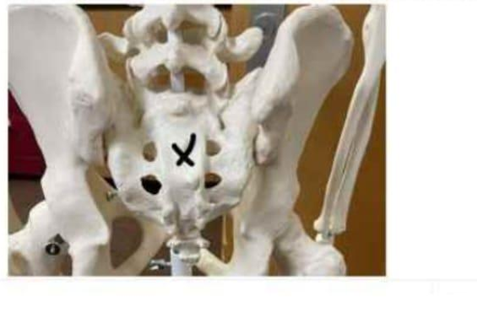
Sacrum
Coccyx
Ilium
Lumbar vertebrae
The Correct Answer is A
The marked structure is the sacrum, a large triangular bone formed by the fusion of five sacral vertebrae located at the base of the vertebral column. It forms the posterior wall of the pelvis and articulates with the ilium at the sacroiliac joints, contributing to pelvic stability and weight transmission from the axial skeleton to the lower limbs. The sacrum also contains sacral foramina that allow passage of spinal nerves.
A. Sacrum: The sacrum is a fused bone consisting of five vertebrae (S1–S5) located between the lumbar spine and coccyx. It forms the posterior portion of the pelvic girdle and articulates with the ilium to distribute body weight during standing and movement. It contains sacral foramina for nerve passage and supports pelvic organs.
B. Coccyx: The coccyx is the small terminal segment of the vertebral column, commonly called the tailbone. It is formed by fusion of 3–5 small vertebrae and lies inferior to the sacrum. It serves as an attachment site for ligaments and pelvic floor muscles. Compared to the sacrum, it is much smaller and more distal, making it unlikely to be the marked structure.
C. Ilium: The ilium is the largest and superior portion of the hip bone, forming the broad flared structure of the pelvis. It articulates with the sacrum at the sacroiliac joint and contributes to the acetabulum. Its main role is weight-bearing and muscle attachment for the abdominal and gluteal muscles. Unlike the midline sacrum, the ilium is lateral and paired on both sides.
D. Lumbar vertebrae: The lumbar vertebrae are five large vertebrae located in the lower back above the sacrum. They provide major support for body weight and allow flexion and extension of the trunk. While they articulate directly with the sacrum, they are separate segmented bones rather than a fused triangular structure.
Nursing Test Bank
Naxlex Comprehensive Predictor Exams
Related Questions
Correct Answer is C
Explanation
The marked structure is the cerebellum, a major part of the hindbrain located posterior to the brainstem and inferior to the occipital lobes of the cerebrum. It consists of two hemispheres connected by the vermis and has a highly folded surface (folia) that increases its cortical area. The cerebellum is primarily responsible for coordination of voluntary movements, maintenance of posture, balance, and fine motor control. It does not initiate movement but ensures that movements are smooth, precise, and well-timed.
A. Cerebrum: The cerebrum is the largest part of the brain and consists of the cerebral hemispheres, including the frontal, parietal, temporal, and occipital lobes. It is responsible for higher cognitive functions such as reasoning, memory, language, and voluntary motor activity. Unlike the cerebellum, it is located superiorly and anteriorly in the cranial cavity and is not involved in fine motor coordination and balance regulation.
B. Brainstem: The brainstem connects the cerebrum and cerebellum to the spinal cord and consists of the midbrain, pons, and medulla oblongata. It regulates vital autonomic functions such as respiration, heart rate, and blood pressure. While it lies close to the cerebellum, it is a vertical structure inferior to the cerebrum rather than a posterior, bilobed structure like the cerebellum.
C. Cerebellum: The cerebellum is located in the posterior cranial fossa, inferior to the occipital lobes and posterior to the brainstem. It is responsible for coordinating voluntary motor activity, maintaining balance, posture, and muscle tone. It receives input from the cerebral cortex and sensory systems to fine-tune motor output. Its highly folded folia and bilateral hemispheres are characteristic features.
D. Medulla oblongata: The medulla oblongata is the lowest part of the brainstem, continuous with the spinal cord. It controls essential autonomic functions such as breathing, heart rate, and blood pressure regulation. Unlike the cerebellum, it is a narrow, tubular structure and does not have a highly folded cortical surface or function in motor coordination and balance.
Correct Answer is B
Explanation
Skeletal muscle contraction depends on excitation–contraction coupling, a process where an electrical signal triggers calcium release, leading to interaction between actin and myosin filaments. Calcium ions are essential because they bind to troponin, shifting tropomyosin away from actin binding sites and allowing cross-bridge formation. The rapid availability of calcium within muscle fibers is ensured by specialized intracellular storage systems. The sarcoplasmic reticulum plays a central role in regulating calcium release and reuptake to control contraction and relaxation.
A. Mitochondria: Mitochondria are organelles responsible for ATP production through aerobic respiration, specifically oxidative phosphorylation. While they are essential for providing energy required for muscle contraction, they do not serve as a primary calcium storage or release site for contraction initiation. Although mitochondria can take up calcium for metabolic regulation, this is not the calcium source that activates troponin.
B. Sarcoplasmic reticulum: the sarcoplasmic reticulum (SR) is the specialized intracellular organelle that stores and releases calcium ions in skeletal muscle fibers. When an action potential travels along the sarcolemma and into the T-tubules, it triggers voltage-sensitive receptors that open calcium channels in the SR. Calcium then floods into the sarcoplasm and binds to troponin C, initiating the contraction process. The SR acts as the primary and rapid regulatory calcium reservoir for muscle excitation–contraction coupling.
C. Extracellular fluid: Extracellular fluid contains calcium ions, but skeletal muscle contraction does not depend on extracellular calcium influx as the primary trigger. Unlike cardiac muscle, skeletal muscle relies mainly on internal calcium stores from the sarcoplasmic reticulum. The small amount of extracellular calcium present is not sufficient or directly responsible for initiating contraction. This source is not the primary contributor to troponin activation in skeletal muscle.
D. T-tubules: T-tubules are invaginations of the sarcolemma that transmit the action potential deep into the muscle fiber. Their role is electrical conduction rather than calcium storage or release. They are structurally linked to the sarcoplasmic reticulum and help trigger calcium release via voltage-sensitive proteins. However, they do not contain or supply calcium themselves, making them an incorrect source of calcium ions for contraction.
Whether you are a student looking to ace your exams or a practicing nurse seeking to enhance your expertise , our nursing education contents will empower you with the confidence and competence to make a difference in the lives of patients and become a respected leader in the healthcare field.
Visit Naxlex, invest in your future and unlock endless possibilities with our unparalleled nursing education contents today