Identify the structure indicated by the arrow.
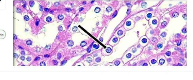
Nucleus
Cytoplasm
Cell membrane
Mitochondria
The Correct Answer is A
In histological staining, such as the hematoxylin and eosin (H&E) stain used in this micrograph, specific cellular structures exhibit distinct staining affinities. Hematoxylin is a basic dye that binds to acidic cellular components, particularly the chromatin within the cell's genetic center, causing it to appear deep blue or purple. Identifying this structure is fundamental for recognizing cellular health, activity levels, and morphological characteristics in various tissue types.
A. The nucleus is the correct identification for the dark, spherical, deep blue-purple structure indicated by the arrow. This organelle contains the cell's genetic material and, due to its high concentration of DNA and acidic proteins, it strongly attracts the hematoxylin stain, making it a prominent landmark in almost all histological tissue sections.
B. The cytoplasm is the gelatinous substance that fills the cell and surrounds the nucleus. In H&E staining, the cytoplasm typically binds to eosin, an acidic dye, and therefore appears various shades of pink or red. The arrow is clearly pointing to the distinct, dark, central body within the cell rather than the surrounding pink-stained area.
C. The cell membrane is the semi-permeable boundary that encapsulates the entire cell. While it is present in the tissue section, it is extremely thin and generally requires special stains or higher magnification to be visualized as a distinct boundary. The arrow is pointing to the large, prominent central organelle, not the delicate outer edge of the cell.
D. Mitochondria are the "powerhouses" of the cell, responsible for ATP production. While they are present within the cytoplasm, they are generally too small to be individually distinguished at this level of magnification without specialized histochemical techniques. They do not appear as large, singular, deep-blue spherical structures like the one indicated by the arrow.
Nursing Test Bank
Naxlex Comprehensive Predictor Exams
Related Questions
Correct Answer is D
Explanation
The marked structure is the parietal lobe, a major division of the cerebral cortex located superiorly in the brain between the frontal and occipital lobes. It lies posterior to the central sulcus and anterior to the occipital lobe, forming a significant portion of the superior and lateral aspects of each cerebral hemisphere. The parietal lobe is primarily responsible for processing somatosensory information such as touch, pressure, temperature, and pain. It also integrates sensory input to support spatial awareness, body orientation, and proprioception.
A. Frontal lobe: The frontal lobe is located anteriorly in the cerebrum, in front of the central sulcus. It is responsible for executive functions such as decision-making, judgment, personality, voluntary motor control, and speech production via Broca’s area. Compared to the parietal lobe, it is more anterior and not primarily involved in somatosensory integration or spatial processing.
B. Temporal lobe: The temporal lobe is located on the lateral aspect of the brain beneath the lateral sulcus. It is primarily involved in auditory processing, language comprehension (Wernicke’s area), and memory formation. Unlike the parietal lobe, it does not process primary somatosensory input or spatial body awareness.
C. Occipital lobe: The occipital lobe is located at the posterior pole of the brain and is the primary center for visual processing. It receives and interprets visual stimuli from the retina via the optic pathways. Compared to the parietal lobe, it is more posterior and specialized for vision rather than somatic sensation or spatial integration.
D. Parietal lobe: The parietal lobe is positioned superiorly and centrally on the cerebral hemispheres, posterior to the frontal lobe and anterior to the occipital lobe. It contains the primary somatosensory cortex located in the postcentral gyrus, which processes tactile and proprioceptive input from the body. It integrates sensory information to support spatial awareness, body positioning, and coordination of movement. Its location and function correspond to the marked region.
Correct Answer is B
Explanation
Skeletal muscle contraction depends on excitation–contraction coupling, a process where an electrical signal triggers calcium release, leading to interaction between actin and myosin filaments. Calcium ions are essential because they bind to troponin, shifting tropomyosin away from actin binding sites and allowing cross-bridge formation. The rapid availability of calcium within muscle fibers is ensured by specialized intracellular storage systems. The sarcoplasmic reticulum plays a central role in regulating calcium release and reuptake to control contraction and relaxation.
A. Mitochondria: Mitochondria are organelles responsible for ATP production through aerobic respiration, specifically oxidative phosphorylation. While they are essential for providing energy required for muscle contraction, they do not serve as a primary calcium storage or release site for contraction initiation. Although mitochondria can take up calcium for metabolic regulation, this is not the calcium source that activates troponin.
B. Sarcoplasmic reticulum: the sarcoplasmic reticulum (SR) is the specialized intracellular organelle that stores and releases calcium ions in skeletal muscle fibers. When an action potential travels along the sarcolemma and into the T-tubules, it triggers voltage-sensitive receptors that open calcium channels in the SR. Calcium then floods into the sarcoplasm and binds to troponin C, initiating the contraction process. The SR acts as the primary and rapid regulatory calcium reservoir for muscle excitation–contraction coupling.
C. Extracellular fluid: Extracellular fluid contains calcium ions, but skeletal muscle contraction does not depend on extracellular calcium influx as the primary trigger. Unlike cardiac muscle, skeletal muscle relies mainly on internal calcium stores from the sarcoplasmic reticulum. The small amount of extracellular calcium present is not sufficient or directly responsible for initiating contraction. This source is not the primary contributor to troponin activation in skeletal muscle.
D. T-tubules: T-tubules are invaginations of the sarcolemma that transmit the action potential deep into the muscle fiber. Their role is electrical conduction rather than calcium storage or release. They are structurally linked to the sarcoplasmic reticulum and help trigger calcium release via voltage-sensitive proteins. However, they do not contain or supply calcium themselves, making them an incorrect source of calcium ions for contraction.
Whether you are a student looking to ace your exams or a practicing nurse seeking to enhance your expertise , our nursing education contents will empower you with the confidence and competence to make a difference in the lives of patients and become a respected leader in the healthcare field.
Visit Naxlex, invest in your future and unlock endless possibilities with our unparalleled nursing education contents today