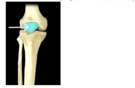
Which structure is highlighted and indicated by the arrow in the image below?
Tibial tuberosity
Femoral condyle
Fibular head
Patella
The Correct Answer is D
The marked structure is the patella, a sesamoid bone embedded within the tendon of the quadriceps femoris muscle group. It is located anterior to the knee joint and articulates with the patellar surface of the femur. The patella increases the mechanical advantage of the quadriceps tendon by improving its angle of pull, which enhances knee extension efficiency. It also protects the anterior aspect of the knee joint from direct trauma.
A. Tibial tuberosity: The tibial tuberosity is a rough anterior projection on the proximal tibia located just inferior to the patella. It serves as the insertion site of the patellar ligament, which connects the patella to the tibia. Unlike the patella, it is part of the tibia and lies below the knee joint rather than within the quadriceps tendon.
B. Femoral condyle: The femoral condyles are the rounded distal projections of the femur that articulate with the tibia and patella to form the knee joint. They are involved in weight transmission and joint movement during flexion and extension. Unlike the patella, they are internal joint surfaces rather than a separate anterior bone.
C. Fibular head: The fibular head is the proximal expanded end of the fibula located on the lateral aspect of the knee. It provides attachment for ligaments and muscles and contributes to lateral knee stability. It is not part of the patellofemoral joint and lies more laterally and posteriorly compared to the patella.
D. Patella: The patella is a triangular sesamoid bone located anterior to the knee joint within the quadriceps tendon. It articulates with the femoral trochlea and enhances the leverage of knee extension by increasing the angle of pull of the quadriceps muscle. It also protects the knee joint from anterior trauma. Since the highlighted structure is a small, superficial bone over the front of the knee joint, it corresponds to the patella.
Nursing Test Bank
Naxlex Comprehensive Predictor Exams
Related Questions
Correct Answer is B
Explanation
Skeletal muscle is organized in a precise hierarchical structure that allows efficient force generation and coordinated movement. This organization begins at the molecular level with contractile proteins and scales up to whole muscle organs. Each level of structure is nested within the next, ensuring proper alignment and transmission of contractile force. Understanding this arrangement is essential for explaining muscle physiology, contraction mechanics, and injury patterns.
A. Muscle → fascicle → muscle fiber → myofibril → filament: This sequence reverses the true anatomical hierarchy. Muscle is the largest structural unit, not the smallest, and should be placed at the end of the progression. Filaments are the smallest contractile elements within myofibrils. This presents a descending order rather than an ascending structural organization.
B. Filament → myofibril → muscle fiber → fascicle → muscle: This is the correct sequence from smallest to largest structural unit in skeletal muscle. Filaments (actin and myosin) form the contractile basis of myofibrils. Myofibrils bundle together to form muscle fibers (cells), which are grouped into fascicles. Fascicles then combine to form the entire skeletal muscle, allowing coordinated contraction and force generation.
C. Filament → muscle fiber → myofibril → fascicle → muscle: This option skips the proper intermediate structural level. Myofibrils exist between filaments and muscle fibers, serving as the contractile units within each muscle cell. Placing muscle fiber before myofibril disrupts the correct cellular organization. This misrepresents the actual histological hierarchy of skeletal muscle.
D. Myofibril → filament → muscle fiber → fascicle → muscle: This sequence places myofibrils above filaments, which reverses their true relationship. Filaments (actin and myosin) are the smallest functional units within myofibrils. Additionally, the correct sequence should begin with filaments, not myofibrils.
Correct Answer is D
Explanation
The eye is composed of the eyeball and several accessory structures that support its function by providing protection, lubrication, and structural maintenance. These accessory structures include the eyelids, conjunctiva, and lacrimal apparatus, which work to preserve the integrity of the ocular surface. However, they do not directly participate in the process of vision. The retina, in contrast, is part of the eyeball itself and is responsible for converting light into electrical impulses for visual processing in the brain.
A. Conjunctiva: The conjunctiva is a thin mucous membrane that lines the inner eyelids and covers the anterior sclera. It contains goblet cells that secrete mucin, contributing to tear film stability and ocular surface lubrication. It also provides immune protection by acting as a barrier against pathogens. Its role is supportive and protective rather than sensory or visual, which classifies it as an accessory structure of the eye.
B. Eyelids: The eyelids are protective anatomical structures composed of skin, muscle, and connective tissue, including the orbicularis oculi and tarsal plates. They function to shield the eye from trauma, regulate light exposure, and distribute tears evenly across the corneal surface during blinking. They also help prevent corneal drying and maintain visual clarity. They support and protect the eye rather than participate in vision, they are considered accessory structures.
C. Lacrimal apparatus: The lacrimal apparatus includes the lacrimal gland, canaliculi, lacrimal sac, and nasolacrimal duct. It is responsible for producing and draining tears that contain water, electrolytes, enzymes such as lysozyme, and immunoglobulins. These components lubricate the eye, nourish the avascular cornea, and provide antimicrobial defense. Because its function is maintenance and protection of the ocular surface rather than visual processing, it is classified as an accessory structure.
D. Retina: The retina is the innermost layer of the eyeball and is derived from neural tissue during embryological development. It contains photoreceptor cells, specifically rods and cones, which convert light energy into electrical signals through phototransduction. These signals are processed by bipolar and ganglion cells, whose axons form the optic nerve. The retina is directly responsible for initiating vision and transmitting visual information to the brain. Therefore, it is not an accessory structure but an essential component of the visual system.
Whether you are a student looking to ace your exams or a practicing nurse seeking to enhance your expertise , our nursing education contents will empower you with the confidence and competence to make a difference in the lives of patients and become a respected leader in the healthcare field.
Visit Naxlex, invest in your future and unlock endless possibilities with our unparalleled nursing education contents today