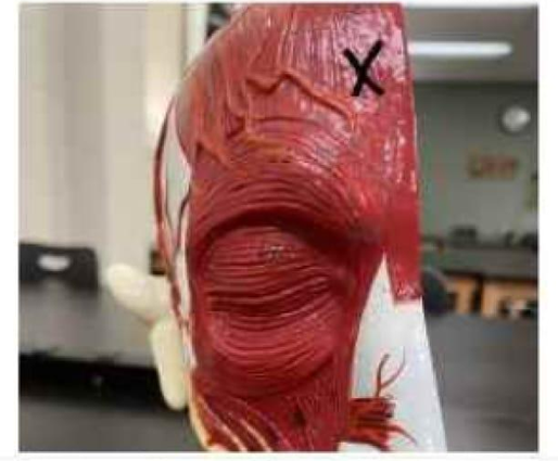
Identify the muscle marked with an X in the image below.
Orbicularis oculi
Masseter
Temporalis
Frontalis
The Correct Answer is D
The marked structure is the frontalis muscle, which is the anterior portion of the occipitofrontalis muscle located in the forehead region. It is a thin, broad muscle of facial expression that lies within the superficial musculoaponeurotic system of the scalp. The frontalis has no bony attachments to the skull; instead, it is anchored to the galea aponeurotica posteriorly. Its primary function is elevation of the eyebrows and wrinkling of the forehead, especially during expressions of surprise or attention.
A. Orbicularis oculi: The orbicularis oculi is a circular muscle surrounding the orbit of the eye. It is responsible for closing the eyelids during blinking and squinting, protecting the eye from debris and excessive light. It has orbital and palpebral portions that control forceful and gentle eyelid closure. Unlike the frontalis, it is located around the eye rather than the forehead region.
B. Masseter: The masseter is a powerful muscle of mastication located on the lateral aspect of the mandible. It elevates the mandible during chewing and is one of the strongest muscles in the body relative to its size. It originates from the zygomatic arch and inserts on the ramus of the mandible. Compared to the frontalis, it is located in the lower face and is involved in jaw movement, not facial expression of the forehead.
C. Temporalis: The temporalis is a broad, fan-shaped muscle located on the lateral side of the skull in the temporal fossa. It assists in elevating and retracting the mandible during chewing. It inserts onto the coronoid process of the mandible and is a key muscle of mastication. Unlike the frontalis, it is deeper, lateral, and involved in jaw movement rather than forehead expression.
D. Frontalis: The frontalis is the frontal belly of the occipitofrontalis muscle, located in the forehead region. It elevates the eyebrows and wrinkles the skin of the forehead, contributing to facial expressions such as surprise and curiosity. It is innervated by the facial nerve (CN VII) and has no direct bony attachment, instead connecting via the epicranial aponeurosis. Since the marked area is in the anterior forehead region, it corresponds to the frontalis muscle.
Nursing Test Bank
Naxlex Comprehensive Predictor Exams
Related Questions
Correct Answer is C
Explanation
Bone formation occurs through two primary processes: intramembranous ossification and endochondral ossification. Intramembranous ossification involves the direct conversion of mesenchymal tissue into bone without a cartilage intermediate. This process is responsible for forming flat bones, especially those of the skull and parts of the clavicle. These bones are crucial for protecting the brain and providing structural support for the head. Understanding bone development is essential for identifying skeletal anatomy and growth patterns.
A. Phalanges of the fingers: The phalanges are formed through endochondral ossification, not intramembranous ossification. In this process, a hyaline cartilage model is first formed and then gradually replaced by bone tissue. This type of ossification is typical of long bones that require elongation and support for movement. Therefore, phalanges are not examples of intramembranous bones.
B. Vertebrae: Vertebrae also develop through endochondral ossification. They begin as cartilage templates that are progressively ossified during fetal development and growth. This process allows for structured shaping and support of the spinal column. Since intramembranous ossification does not involve a cartilage stage, vertebrae are not classified as intramembranous bones.
C. Bones of the cranium: most cranial bones (such as the frontal, parietal, and portions of the occipital bones) are formed through intramembranous ossification. In this process, mesenchymal cells directly differentiate into osteoblasts, which lay down bone matrix without a cartilage precursor. This allows for the formation of flat, protective bones of the skull that safeguard the brain.
D. Femur: The femur is a long bone that develops through endochondral ossification. It begins as a cartilage model that is gradually replaced by bone tissue during growth. This process supports longitudinal growth and structural strength needed for weight-bearing and locomotion. Therefore, it is not an intramembranous bone.
Correct Answer is D
Explanation
The marked structure is the frontalis muscle, which is the anterior portion of the occipitofrontalis muscle located in the forehead region. It is a thin, broad muscle of facial expression that lies within the superficial musculoaponeurotic system of the scalp. The frontalis has no bony attachments to the skull; instead, it is anchored to the galea aponeurotica posteriorly. Its primary function is elevation of the eyebrows and wrinkling of the forehead, especially during expressions of surprise or attention.
A. Orbicularis oculi: The orbicularis oculi is a circular muscle surrounding the orbit of the eye. It is responsible for closing the eyelids during blinking and squinting, protecting the eye from debris and excessive light. It has orbital and palpebral portions that control forceful and gentle eyelid closure. Unlike the frontalis, it is located around the eye rather than the forehead region.
B. Masseter: The masseter is a powerful muscle of mastication located on the lateral aspect of the mandible. It elevates the mandible during chewing and is one of the strongest muscles in the body relative to its size. It originates from the zygomatic arch and inserts on the ramus of the mandible. Compared to the frontalis, it is located in the lower face and is involved in jaw movement, not facial expression of the forehead.
C. Temporalis: The temporalis is a broad, fan-shaped muscle located on the lateral side of the skull in the temporal fossa. It assists in elevating and retracting the mandible during chewing. It inserts onto the coronoid process of the mandible and is a key muscle of mastication. Unlike the frontalis, it is deeper, lateral, and involved in jaw movement rather than forehead expression.
D. Frontalis: The frontalis is the frontal belly of the occipitofrontalis muscle, located in the forehead region. It elevates the eyebrows and wrinkles the skin of the forehead, contributing to facial expressions such as surprise and curiosity. It is innervated by the facial nerve (CN VII) and has no direct bony attachment, instead connecting via the epicranial aponeurosis. Since the marked area is in the anterior forehead region, it corresponds to the frontalis muscle.
Whether you are a student looking to ace your exams or a practicing nurse seeking to enhance your expertise , our nursing education contents will empower you with the confidence and competence to make a difference in the lives of patients and become a respected leader in the healthcare field.
Visit Naxlex, invest in your future and unlock endless possibilities with our unparalleled nursing education contents today