Identify the muscle that is highlighted and circled in the image below of the posterior side of a huma body.
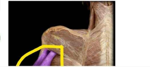
Latissimus dorsi
Triceps brachii
Deltoid (posterior fibers)
Biceps brachii
The Correct Answer is B
The marked structure is the triceps brachii muscle, the primary extensor of the elbow joint located in the posterior compartment of the upper arm. It consists of three heads: long head (originating from the infraglenoid tubercle of the scapula), lateral head, and medial head (both originating from the posterior humerus). All three heads converge to insert on the olecranon process of the ulna. The triceps brachii is responsible for elbow extension and assists in shoulder stabilization, particularly through the long head.
A. Latissimus dorsi: The latissimus dorsi is a large, flat, triangular muscle of the lower back that extends to the humerus. It functions in shoulder extension, adduction, and internal rotation, especially during pulling movements. While it is visible in posterior views, it is located inferiorly and medially on the back rather than forming the posterior upper arm mass. It does not act as the primary extensor of the elbow joint.
B. Triceps brachii: The triceps brachii is the main muscle of the posterior upper arm, occupying most of its surface area. It has three heads that originate from the scapula and humerus and converge onto the olecranon of the ulna. Its primary function is extension of the forearm at the elbow joint, especially during pushing actions such as pushing open a door or extending the arm against resistance. The long head also assists in shoulder extension and stabilization, making it the correct answer for a posterior upper arm highlight.
C. Deltoid (posterior fibers): The posterior fibers of the deltoid are located on the lateral shoulder and contribute to shoulder extension, horizontal abduction, and external rotation of the arm. Although visible from a posterior view, they are situated over the shoulder joint rather than the posterior upper arm. They do not extend the elbow joint, which distinguishes them from the triceps brachii.
D. Biceps brachii: The biceps brachii is located in the anterior compartment of the upper arm and is primarily responsible for elbow flexion and forearm supination. It is not visible as a dominant posterior muscle and does not contribute to the posterior contour of the upper arm. Its anatomical position on the front of the arm makes it distinct from the triceps brachii.
Nursing Test Bank
Naxlex Comprehensive Predictor Exams
Related Questions
Correct Answer is C
Explanation
The marked structure is the carpals, a group of eight small bones that form the wrist (carpus). These bones are arranged in two rows and create the connection between the forearm and the hand. The carpals provide flexibility and stability to the wrist joint, allowing a wide range of movements including flexion, extension, abduction, and adduction. They also play a key role in absorbing and distributing forces transmitted from the hand to the forearm during gripping and weight-bearing activities.
A. Metacarpals: The metacarpals are five long bones located in the palm of the hand, positioned between the carpals and the phalanges. They form the framework of the hand and support finger movements and grip strength. Compared to the carpals, they are longer and more tubular, forming the “palm bones” rather than the wrist structure.
B. Radius: The radius is the lateral forearm bone on the thumb side that extends from the elbow to the wrist. It articulates distally with the carpals to form part of the wrist joint but is not part of the wrist bones themselves. Unlike the carpals, it is a long bone of the forearm rather than a cluster of small bones.
C. Carpals: The carpals are eight small, irregular bones forming the wrist joint between the forearm and the hand. They are arranged in proximal and distal rows and provide both stability and flexibility to wrist movements. Their function includes shock absorption during hand impact and facilitating smooth movement between the forearm and hand. Since the circled structure is a compact cluster at the wrist, it corresponds to the carpals.
D. Phalanges: The phalanges are the bones of the fingers and thumb, arranged in three segments per finger (except the thumb, which has two). They allow fine motor movements such as grasping and writing. Compared to the carpals, they are distal and elongated structures forming the digits rather than the wrist joint itself.
Correct Answer is A
Explanation
The marked structure is the trapezius muscle, a large, superficial, triangular muscle that extends across the posterior neck and upper back. It originates from the occipital bone, ligamentum nuchae, and spinous processes of C7–T12, and inserts onto the clavicle, acromion, and spine of the scapula. The trapezius plays a major role in scapular positioning and movement, including elevation, retraction, depression, and upward rotation. It also contributes to neck extension and stabilization of the shoulder girdle.
A. Trapezius: The trapezius is a broad, superficial muscle covering the posterior neck and upper thorax, forming a diamond-shaped structure across the upper back. It controls scapular movements such as elevation (shrugging), retraction (pulling shoulders back), and rotation necessary for overhead arm activity. It is also involved in stabilizing the scapula during upper limb movement. Its extensive posterior location and attachment to the scapula and clavicle make it the correct answer.
B. Latissimus dorsi: The latissimus dorsi is a large, flat muscle of the lower back that extends to the humerus. It functions primarily in shoulder extension, adduction, and internal rotation, especially during pulling movements. Unlike the trapezius, it is located in the lower posterior trunk and does not extend into the neck region. It also does not elevate or stabilize the scapula in the same way.
C. Deltoid: The deltoid is a thick, triangular muscle covering the lateral aspect of the shoulder joint. It is responsible for abduction of the arm and contributes to flexion and extension depending on fiber segment. It is not located on the posterior back or neck, and it does not control scapular movement, unlike the trapezius.
D. Sternocleidomastoid: The sternocleidomastoid is a paired muscle located in the anterior and lateral neck. It originates from the sternum and clavicle and inserts on the mastoid process of the temporal bone. It functions in neck flexion, rotation, and lateral bending. Compared to the trapezius, it is anteriorly positioned and does not act on the scapula or upper back region.
Whether you are a student looking to ace your exams or a practicing nurse seeking to enhance your expertise , our nursing education contents will empower you with the confidence and competence to make a difference in the lives of patients and become a respected leader in the healthcare field.
Visit Naxlex, invest in your future and unlock endless possibilities with our unparalleled nursing education contents today