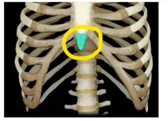
Identify the structure that is circled and highlighted.
Manubrium
Body of sternum
Xiphoid process
Costal cartilage
The Correct Answer is C
The marked structure is the xiphoid process, the smallest and most inferior portion of the sternum. It is a thin, elongated structure located below the body of the sternum and serves as an important anatomical landmark in the thorax. During early life, the xiphoid process is composed primarily of hyaline cartilage and gradually ossifies with age. It provides attachment sites for the diaphragm, rectus abdominis, and transversus thoracis muscles, contributing to respiration, trunk movement, and stabilization of the anterior thoracic wall.
A. Manubrium: The manubrium is the broad, superior portion of the sternum that articulates with the clavicles and the first pair of ribs. It contains the jugular notch and clavicular notches, making it an important landmark for identifying thoracic structures. Its primary function is to provide attachment and support for the pectoral girdle and upper ribs. Unlike the xiphoid process, it is located at the superior end of the sternum rather than the inferior tip.
B. Body of sternum: The body of the sternum is the largest and longest portion of the sternum, situated between the manubrium and xiphoid process. It articulates with the costal cartilages of ribs 2 through 7 and contributes significantly to protection of the heart, lungs, and major thoracic vessels. Compared with the xiphoid process, it is much larger and forms the central portion of the anterior thoracic cage.
C. Xiphoid process: The xiphoid process is the small, inferior segment of the sternum located immediately below the sternal body. It serves as an attachment point for the diaphragm, rectus abdominis, and transversus thoracis muscles, making it important in respiration and trunk stabilization. Clinically, it is used as a landmark during cardiopulmonary resuscitation (CPR), as incorrect hand placement over the xiphoid can result in fracture and injury to underlying organs. Since the highlighted structure is the inferior terminal part of the sternum, it corresponds to the xiphoid process.
D. Costal cartilage: Costal cartilage consists of bars of hyaline cartilage that connect the anterior ends of the ribs to the sternum. These cartilages provide elasticity to the thoracic cage, allowing expansion and recoil during breathing. They are paired structures extending laterally from the sternum rather than a single midline structure. Unlike the xiphoid process, costal cartilage is not a component of the sternum itself but serves as a connection between ribs and sternum.
Nursing Test Bank
Naxlex Comprehensive Predictor Exams
Related Questions
Correct Answer is A
Explanation
The vertebra is a complex irregular bone designed to provide structural support, flexibility, and protection for the spinal cord. It consists of a central body, a vertebral arch, and several specialized bony projections that serve as attachment points for muscles and ligaments. In the image, the structure marked with an "X" is one of the lateral projections extending from the vertebral arch, which is essential for stabilizing the vertebral column and facilitating movement through muscular pull.
A. The transverse process is the lateral bony projection that extends from the junction of the pedicle and lamina on each side of the vertebra. These processes serve as critical attachment sites for deep back muscles and, in the thoracic region, articulate with the ribs. The structure marked with an "X" is clearly identified as this lateral projection.
B. The spinous process is the singular, posterior-facing projection located at the midline of the vertebral arch. It is easily palpable through the skin along the back and serves as an anchor for various ligaments and muscles that support the spine. It is distinct from the lateral structure marked in the image, which originates from the side of the arch.
C. The superior articular facet is a smooth surface on the superior aspect of the vertebral arch that articulates with the inferior articular facet of the vertebra above it. These facets are crucial for limiting movement and maintaining the alignment of the vertebral column. They are located near the pedicle-lamina junction but are not the lateral extension identified by the "X" in the image.
D. The vertebral foramen is the large, central opening created by the vertebral arch and the posterior surface of the vertebral body. It houses the spinal cord, its associated protective meninges, and the surrounding blood vessels. The mark "X" is placed on the external lateral surface of the bone, not within this central canal.
Correct Answer is A
Explanation
Muscle tissue in the human body is classified into three types based on structure and control mechanisms: skeletal, smooth, and cardiac muscle. These tissues differ in their microscopic organization, location, and mode of nervous system regulation. Voluntary control refers to conscious activation via the somatic nervous system, while involuntary control is regulated by the autonomic nervous system and intrinsic pacemaker activity. Understanding these differences is essential for distinguishing body systems involved in movement versus automatic physiological functions.
A. Smooth muscle tissue and cardiac muscle tissue: both smooth and cardiac muscle tissues are under involuntary control. Smooth muscle, found in structures such as blood vessels, the gastrointestinal tract, and respiratory passages, is regulated by the autonomic nervous system and local chemical signals. Cardiac muscle, located in the heart, is also involuntary and has intrinsic rhythmic activity controlled by the sinoatrial node, with modulation from autonomic inputs. Both muscle types function automatically without conscious control.
B. Skeletal muscle tissue and smooth muscle tissue: skeletal muscle is under voluntary control, not involuntary control. Skeletal muscles are innervated by the somatic nervous system and require conscious effort for activation, such as walking or lifting objects. While smooth muscle is involuntary, pairing it with skeletal muscle makes this option incorrect overall.
C. Smooth muscle tissue, skeletal muscle tissue, and cardiac muscle tissue: This option includes all three muscle types, but skeletal muscle is voluntary. Although smooth and cardiac muscles are involuntary, skeletal muscle is consciously controlled. Therefore, not all listed muscle types are involuntary.
D. Skeletal muscle tissue only: skeletal muscle is entirely voluntary and controlled by the somatic nervous system. It is responsible for conscious movements such as locomotion and posture maintenance. It does not function automatically or independently of conscious control. Therefore, it cannot be classified as involuntary muscle tissue.
Whether you are a student looking to ace your exams or a practicing nurse seeking to enhance your expertise , our nursing education contents will empower you with the confidence and competence to make a difference in the lives of patients and become a respected leader in the healthcare field.
Visit Naxlex, invest in your future and unlock endless possibilities with our unparalleled nursing education contents today