Identify the structure circled in the image below.
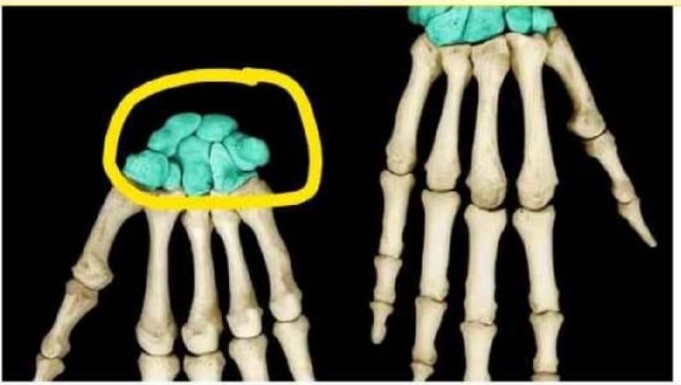
Metacarpals
Radius
Carpals
Phalanges
The Correct Answer is C
The marked structure is the carpals, a group of eight small bones that form the wrist (carpus). These bones are arranged in two rows and create the connection between the forearm and the hand. The carpals provide flexibility and stability to the wrist joint, allowing a wide range of movements including flexion, extension, abduction, and adduction. They also play a key role in absorbing and distributing forces transmitted from the hand to the forearm during gripping and weight-bearing activities.
A. Metacarpals: The metacarpals are five long bones located in the palm of the hand, positioned between the carpals and the phalanges. They form the framework of the hand and support finger movements and grip strength. Compared to the carpals, they are longer and more tubular, forming the “palm bones” rather than the wrist structure.
B. Radius: The radius is the lateral forearm bone on the thumb side that extends from the elbow to the wrist. It articulates distally with the carpals to form part of the wrist joint but is not part of the wrist bones themselves. Unlike the carpals, it is a long bone of the forearm rather than a cluster of small bones.
C. Carpals: The carpals are eight small, irregular bones forming the wrist joint between the forearm and the hand. They are arranged in proximal and distal rows and provide both stability and flexibility to wrist movements. Their function includes shock absorption during hand impact and facilitating smooth movement between the forearm and hand. Since the circled structure is a compact cluster at the wrist, it corresponds to the carpals.
D. Phalanges: The phalanges are the bones of the fingers and thumb, arranged in three segments per finger (except the thumb, which has two). They allow fine motor movements such as grasping and writing. Compared to the carpals, they are distal and elongated structures forming the digits rather than the wrist joint itself.
Nursing Test Bank
Naxlex Comprehensive Predictor Exams
Related Questions
Correct Answer is A
Explanation
The skin is composed of three main layers: the epidermis, dermis, and hypodermis (subcutaneous layer). The epidermis is the outermost layer and is responsible for forming a protective barrier against environmental damage. It undergoes continuous renewal through a process called keratinization, where cells move from the basal layer to the surface and are eventually shed. Disorders affecting desquamation, such as ichthyosis, primarily involve abnormalities in this outer epithelial layer, leading to accumulation of dead keratinized cells and scaly skin.
A. Epidermis: ichthyosis affects the epidermis, specifically the stratum corneum, which is the outermost portion of the epidermis. Normally, keratinocytes undergo a regulated process of differentiation and desquamation, where dead cells are shed from the skin surface. In ichthyosis, this process is disrupted, leading to excessive accumulation of keratinized cells and a thick, scaly appearance. The epidermis is responsible for barrier function and continuous renewal, making it the primary site of pathology.
B. Dermis: The dermis is the deeper layer of the skin located beneath the epidermis and is composed of connective tissue containing collagen, elastin, blood vessels, nerves, and hair follicles. It provides structural support, elasticity, and nourishment to the epidermis. While it plays an important supportive role, it is not involved in keratinization or surface cell shedding.
C. Hypodermis: The hypodermis, also known as the subcutaneous layer, is the deepest layer of the skin and is primarily composed of adipose tissue and loose connective tissue. It functions in insulation, energy storage, and cushioning of underlying structures. It does not participate in epidermal cell turnover or keratinization. As a result, it is not involved in the pathological process seen in ichthyosis.
D. Subcutaneous layer: The subcutaneous layer is another term for the hypodermis and shares the same structure and functions. It lies beneath the dermis and consists mainly of fat and connective tissue. Its role is primarily supportive and metabolic rather than epithelial renewal. Since ichthyosis is a disorder of epidermal desquamation, the subcutaneous layer is not involved in this condition.
Correct Answer is A
Explanation
Skeletal muscle contraction is based on the sliding filament theory, where thin (actin) and thick (myosin) filaments interact to produce force and movement. These interactions occur in a highly organized structural unit within myofibrils. The arrangement of sarcomeres in series allows coordinated shortening of muscle fibers. Understanding the functional unit of contraction is essential for explaining how muscles generate tension at the microscopic level.
A. Sarcomere: The sarcomere is the correct answer because it is the smallest functional (contractile) unit of skeletal muscle. It is defined as the segment between two Z-discs and contains organized actin and myosin filaments. During contraction, myosin heads bind to actin and pull the thin filaments inward, shortening the sarcomere. This coordinated shortening of many sarcomeres produces overall muscle contraction.
B. Myosin cross-bridge: a cross-bridge is a molecular interaction, not a complete functional unit. It refers specifically to the temporary attachment between a myosin head and an actin binding site. While cross-bridge cycling generates force, it occurs within the sarcomere and depends on its structural organization. It is a mechanism within the functional unit rather than the unit itself.
C. Muscle fiber: a muscle fiber is a single multinucleated muscle cell containing many myofibrils. Although it is the cellular level at which contraction occurs, it is not the smallest functional unit. Each muscle fiber contains thousands of sarcomeres arranged in series and parallel. Contraction occurs within the fiber, but the sarcomere is the true functional unit.
D. Myofibril: a myofibril is a long cylindrical structure within a muscle fiber composed of repeating sarcomeres. It serves as the structural framework for contraction but is not itself the basic contractile unit. Myofibrils transmit force generated by sarcomeres along the length of the muscle cell. It is an organizational structure rather than the functional unit of contraction.
Whether you are a student looking to ace your exams or a practicing nurse seeking to enhance your expertise , our nursing education contents will empower you with the confidence and competence to make a difference in the lives of patients and become a respected leader in the healthcare field.
Visit Naxlex, invest in your future and unlock endless possibilities with our unparalleled nursing education contents today