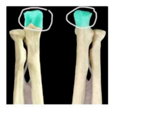
Which structure is highlighted and circled in the image of the lower arm bones below?
Radial head
Coronoid process of ulna
Olecranon process of ulna
Trochlea of humerus
The Correct Answer is C
The marked structure is the olecranon process of the ulna, which is the large, proximal bony prominence of the ulna forming the tip of the elbow. It projects posteriorly and fits into the olecranon fossa of the humerus during elbow extension, stabilizing the elbow joint. The ulna is the medial forearm bone and plays a key role in forming a stable hinge joint at the elbow with the humerus, allowing flexion and extension movements.
A. Radial head: The radial head is the proximal, disc-shaped end of the radius located laterally in the forearm. It articulates with the capitulum of the humerus and the radial notch of the ulna, allowing pronation and supination of the forearm. Unlike the olecranon, it is smooth and rounded rather than a large posterior projection of the elbow.
B. Coronoid process of ulna: The coronoid process is a triangular anterior projection of the proximal ulna. It fits into the coronoid fossa of the humerus during elbow flexion, preventing anterior dislocation. Compared to the olecranon, it is anterior and inferior rather than the prominent posterior elbow tip.
C. Olecranon process of ulna: The olecranon process is the large, posterior bony prominence of the proximal ulna that forms the tip of the elbow. It inserts into the olecranon fossa of the humerus during extension, acting as a lever for triceps brachii muscle attachment. It is easily palpable at the back of the elbow and is the main structure responsible for elbow extension mechanics.
D. Trochlea of humerus: The trochlea is a spool-shaped articular surface located on the distal humerus medially. It articulates with the ulna (specifically the trochlear notch) to form the hinge of the elbow joint. Unlike the olecranon process, it is part of the humerus rather than the ulna and is not a projecting bony landmark at the elbow tip.
Nursing Test Bank
Naxlex Comprehensive Predictor Exams
Related Questions
Correct Answer is C
Explanation
The human skeleton is divided into two major parts: the axial skeleton and the appendicular skeleton. The axial skeleton forms the central axis of the body and is responsible for supporting the head, neck, and trunk. It also provides protection for vital organs such as the brain, spinal cord, and thoracic organs. Understanding this division is essential for identifying bone groups and their functional roles in movement, support, and protection.
A. Radius, ulna, carpals, and phalanges: These bones are part of the upper limb and therefore belong to the appendicular skeleton. The radius and ulna form the forearm, while the carpals and phalanges make up the wrist and fingers. Their primary function is to facilitate movement and manipulation of objects. Since they are located in the limbs rather than the central body axis, they are not part of the axial skeleton.
B. Femur, tibia, fibula, and patella: These bones belong to the lower limb and are part of the appendicular skeleton. The femur is the thigh bone, the tibia and fibula form the lower leg, and the patella is the kneecap. Together, they support weight-bearing and locomotion. However, they are not part of the central axis of the body, so they are excluded from the axial skeleton.
C. Skull, hyoid bone, thoracic cage, and vertebral column: these structures form the axial skeleton. The skull protects the brain, the vertebral column houses the spinal cord, the thoracic cage (ribs and sternum) protects the heart and lungs, and the hyoid bone supports tongue and swallowing functions. Collectively, these structures form the central framework of the body and provide protection and support for vital organs.
D. Scapula, clavicle, humerus, and pelvic bones: These bones are part of the appendicular skeleton, which includes the girdles and limbs. The scapula and clavicle form the shoulder girdle, the humerus is the upper arm bone, and the pelvic bones support the lower trunk and connect the lower limbs to the axial skeleton. Their primary role is movement and attachment of limbs rather than central body support, so they are not part of the axial skeleton.
Correct Answer is D
Explanation
The marked structure is the lens, a transparent, biconvex, avascular structure located posterior to the iris and anterior to the vitreous body. It is suspended by zonular fibers (suspensory ligaments) attached to the ciliary body. The lens plays a critical role in vision by providing fine focusing of light onto the retina through the process of accommodation. Its curvature changes depending on whether the eye is focusing on near or distant objects, allowing precise image formation.
A. Cornea: The cornea is the transparent, dome-shaped anterior surface of the eye that provides most of the eye’s refractive power. It is the first structure that light passes through and contributes significantly to bending light toward the retina. Unlike the lens, it is external, fixed in shape, and continuous with the sclera, serving both protective and optical roles.
B. Retina: The retina is the innermost neural layer lining the posterior aspect of the eye. It contains photoreceptor cells (rods and cones) that convert light into electrical signals sent via the optic nerve to the brain. Unlike the lens, it does not focus light but instead processes visual information after image formation occurs.
C. Iris: The iris is the pigmented muscular structure located anterior to the lens and posterior to the cornea. It regulates pupil size to control the amount of light entering the eye through contraction and relaxation of its smooth muscles. Unlike the lens, it does not contribute to focusing light but only regulates light entry.
D. Lens: The lens is a transparent, flexible, biconvex structure located directly behind the iris. It fine-tunes the focusing of light rays onto the retina through accommodation, changing its curvature via the ciliary muscles. It plays a key role in sharp image formation at varying distances. Its central posterior position relative to the iris makes it the correct structure.
Whether you are a student looking to ace your exams or a practicing nurse seeking to enhance your expertise , our nursing education contents will empower you with the confidence and competence to make a difference in the lives of patients and become a respected leader in the healthcare field.
Visit Naxlex, invest in your future and unlock endless possibilities with our unparalleled nursing education contents today