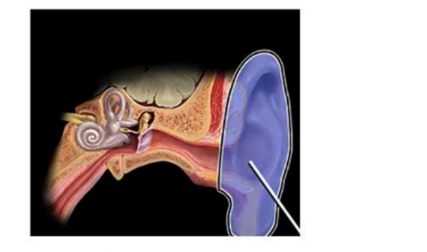
Identify the structure marked below
Tympanic membrane
External auditory canal
Pinna (auricle)
Cochlea
The Correct Answer is C
The marked structure is the pinna (auricle), the visible external part of the ear composed of elastic cartilage covered by skin. It forms the most lateral component of the external ear and is responsible for collecting and directing sound waves into the external auditory canal. Its characteristic folds (helix, antihelix, concha, tragus) help in sound localization by modifying sound wave direction and frequency filtering before transmission to the tympanic membrane. It plays an important role in auditory spatial awareness.
A. Tympanic membrane: The tympanic membrane (eardrum) is a thin, semi-transparent membrane located at the end of the external auditory canal. It vibrates in response to sound waves and transmits mechanical energy to the ossicles of the middle ear. Unlike the pinna, it is not externally visible and lies deep within the external ear canal, separating the external and middle ear.
B. External auditory canal: The external auditory canal is a tubular passage that extends from the pinna to the tympanic membrane. It is lined with skin containing ceruminous glands that produce earwax for protection. Its function is to conduct and slightly amplify sound waves toward the eardrum. Compared to the pinna, it is a deep canal rather than an external visible structure.
C. Pinna (auricle): The pinna is the external, cartilaginous portion of the ear that is visible on the side of the head. It functions to collect sound waves and funnel them into the external auditory canal while also aiding in sound localization by altering sound wave direction. Its unique ridges and depressions help differentiate sounds coming from different directions. Because it is the most external ear structure shown, it is the correct answer.
D. Cochlea: The cochlea is a spiral-shaped structure located in the inner ear within the temporal bone. It contains the organ of Corti, which converts mechanical sound vibrations into electrical nerve impulses. Unlike the pinna, it is deeply embedded within the skull and is responsible for hearing transduction rather than sound collection.
Nursing Test Bank
Naxlex Comprehensive Predictor Exams
Related Questions
Correct Answer is B
Explanation
Metabolism refers to the totality of biochemical processes that occur within living organisms to maintain life. These processes include both anabolic reactions, which build complex molecules from simpler ones, and catabolic reactions, which break down molecules to release energy. Metabolism is essential for growth, repair, reproduction, and the maintenance of cellular functions. It is a continuous and highly regulated process that ensures cells and tissues receive the energy and substrates required for survival.
A. The removal of wastes produced by chemical reactions: This describes excretion rather than metabolism. Excretion involves eliminating metabolic waste products such as carbon dioxide, urea, and ammonia from the body. While waste removal is a consequence of metabolic activity, it is not the definition of metabolism itself. Therefore, this option represents only a small component of physiological function rather than the full concept of metabolism.
B. The chemical reactions occurring in an organism that support life: metabolism includes all chemical reactions within a living organism that sustain life. These reactions involve energy production (catabolism) and biosynthesis (anabolism), both of which are essential for maintaining cellular structure and function. Metabolism encompasses processes such as respiration, protein synthesis, and nutrient breakdown. Together, these reactions ensure survival, growth, and homeostasis.
C. The breakdown of food only during digestion: This limits metabolism to digestion alone. While digestion is part of catabolic metabolism, metabolism also includes cellular respiration, biosynthesis, and energy storage processes. Additionally, metabolic reactions occur at the cellular level, not just within the digestive tract. Therefore, this definition is too narrow and does not represent the full scope of metabolism.
D. The transport of oxygen throughout the body: This option describes a function of the circulatory and respiratory systems rather than metabolism. Oxygen transport is carried out by red blood cells and the cardiovascular system to support cellular respiration. While oxygen is essential for metabolic reactions, its transport is not itself metabolism.
Correct Answer is C
Explanation
The eye is a highly specialized sensory organ composed of refractive media and supportive structures that work together to focus light onto the retina. The lens must be precisely positioned and dynamically adjusted to allow accommodation for near and distant vision. Its stability and shape are maintained by suspensory structures located in the middle vascular layer of the eye. These structures play a crucial role in anchoring the lens while allowing controlled changes in curvature during focusing.
A. Iris: The iris is a pigmented, circular muscular structure located anterior to the lens and surrounding the pupil. It regulates the amount of light entering the eye by controlling pupil diameter through contraction and relaxation of smooth muscle fibers. Although it lies close to the lens, it does not attach to or support the lens structurally. Its function is light regulation rather than mechanical suspension.
B. Cornea: The cornea is the transparent, avascular anterior surface of the eye that provides most of the eye’s refractive power. It serves as the primary entry point for light and contributes significantly to focusing images onto the retina. Anatomically, it is located anterior to the aqueous humor and is completely separate from the lens and its supporting structures. It has no role in anchoring or stabilizing the lens.
C. Ciliary body (ciliary muscle and zonule): The ciliary body is part of the uveal (middle vascular) layer of the eye and consists of the ciliary muscle and ciliary processes. The zonular fibers (suspensory ligaments of the lens) extend from the ciliary processes and attach directly to the lens capsule. These fibers physically suspend the lens in position within the posterior chamber. The ciliary muscle adjusts tension on the zonules to change lens curvature during accommodation, making this structure essential for both lens positioning and focusing.
D. Retina: The retina is the innermost neural layer of the eye responsible for phototransduction, where rods and cones convert light into electrical signals. It lines the posterior segment of the eyeball and connects to the optic nerve. While it is essential for vision, it is anatomically located at the back of the eye and has no structural role in supporting or holding the lens. Its function is sensory rather than mechanical.
Whether you are a student looking to ace your exams or a practicing nurse seeking to enhance your expertise , our nursing education contents will empower you with the confidence and competence to make a difference in the lives of patients and become a respected leader in the healthcare field.
Visit Naxlex, invest in your future and unlock endless possibilities with our unparalleled nursing education contents today