After traveling through the AV node, an electrical impulse travel to which of the following components of an effective cardiac conduction system?
Purkinje fibers
Bundle of His
Bundle branches
SA node
The Correct Answer is B
a. Purkinje fibers: Purkinje fibers are the last structures to receive the signal within the ventricles, causing them to contract.
b. Bundle of His: The correct sequence of the cardiac conduction system is as follows: The electrical impulse originates in the sinoatrial (SA) node, then travels to the atrioventricular (AV) node. From the AV node, the impulse travels to the Bundle of His. After the Bundle of His, the impulse travels through the bundle branches and finally reaches the Purkinje fibers, which facilitate the contraction of the ventricles.
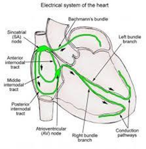
c. Bundle branches: The electrical impulse for heartbeat originates in the SA node (sinoatrial node). It then travels to the AV node (atrioventricular node), which delays the signal before sending it to the Bundle of His. The Bundle of His splits into right and left bundle branches, which distribute the electrical signal to the Purkinje fibers in the ventricles, causing them to contract in a coordinated fashion.
d. SA node: The SA node initiates the electrical impulse, not receive it after the AV node.
Nursing Test Bank
Naxlex Comprehensive Predictor Exams
Related Questions
Correct Answer is B
Explanation
a. Destroy pathogens that enter a break in the skin: While mast cells can contribute to the defense against pathogens by releasing inflammatory mediators, their primary function is not the direct destruction of pathogens.
b. Produce histamine and leukotrienes that initiate inflammation: This is correct. Mast cells are known for their role in releasing histamine and leukotrienes, which are key mediators of the inflammatory response.
c. Cushion bony prominences: This is not a function of mast cells. Cushioning bony prominences is a role more related to adipose tissue.
d. Connect skin to muscles: This is the function of connective tissue, not mast cells.
Correct Answer is B
Explanation
a: Testes - The testes are located outside the body cavity within the scrotum and are responsible for producing sperm and testosterone.
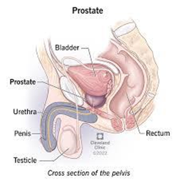
b. Prostate: The prostate gland is a male reproductive organ located at the neck of the bladder and surrounds the urethra. It plays a role in the production of seminal fluid, which nourishes and transports sperm.
c: Rugae - Rugae are folds in the mucous membrane lining the urinary bladder that allow it to stretch as it fills with urine; they do not surround the urethra.
d: Bulbourethral - The bulbourethral glands (Cowper's glands) are pea-sized glands located below the prostate gland that secrete a clear fluid into the urethra during sexual arousal, but they do not surround the urethra at the neck of the bladder.
Whether you are a student looking to ace your exams or a practicing nurse seeking to enhance your expertise , our nursing education contents will empower you with the confidence and competence to make a difference in the lives of patients and become a respected leader in the healthcare field.
Visit Naxlex, invest in your future and unlock endless possibilities with our unparalleled nursing education contents today