Which structural feature of the heart is responsible for preventing the backflow of blood from the left ventricles to the left atrium?
Mitral valve
Tricuspid valve
Aortic valve
Pulmonary valve
The Correct Answer is A
Choice A reason: The mitral valve, also known as the bicuspid valve, is an atrioventricular valve located between the left atrium and the left ventricle. Its primary physiological function is to ensure unidirectional blood flow by closing tightly during ventricular systole, thereby preventing regurgitation of high-pressure blood back into the left atrium.
Choice B reason: The tricuspid valve is situated between the right atrium and the right ventricle. It functions to prevent the retrograde flow of deoxygenated blood into the right atrium during right ventricular contraction. It is anatomically positioned on the right side of the heart and does not manage left-sided pressures.
Choice C reason: The aortic valve is a semilunar valve located between the left ventricle and the aorta. Its function is to prevent blood from flowing back from the systemic circulation into the left ventricle during ventricular diastole. It does not regulate blood flow between the atrial and ventricular chambers.
Choice D reason: The pulmonary valve is a semilunar valve positioned between the right ventricle and the pulmonary artery. It serves to prevent the backflow of deoxygenated blood from the pulmonary vasculature into the right ventricle during the relaxation phase of the cardiac cycle, maintaining efficient forward flow to the lungs.
Nursing Test Bank
Naxlex Comprehensive Predictor Exams
Related Questions
Correct Answer is B
Explanation
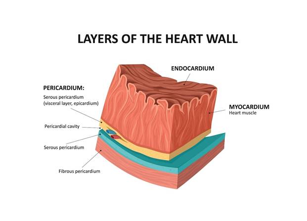
Choice A reason: The endocardium is the innermost layer of the heart, consisting of endothelial cells that line the chambers and valves. While it provides a smooth, frictionless surface for blood flow, it does not possess the contractile properties necessary to generate the mechanical force required for pumping blood.
Choice B reason: The myocardium is the thick, muscular middle layer of the heart wall. It is composed of specialized cardiac muscle cells that possess the contractile ability to generate high pressure and force, making it the primary engine responsible for the mechanical pumping action that circulates blood throughout the body.
Shutterstock
Choice C reason: The epicardium is the outer serous layer of the heart wall, also known as the visceral layer of the serous pericardium. It serves a protective function and contains coronary blood vessels and adipose tissue, but it plays no role in the direct contractile force production for blood circulation.
Choice D reason: The pericardium is the fibrous sac that surrounds and protects the heart. It provides physical support and prevents the heart from over-expanding. While it is essential for cardiac health, it is a structural covering and does not contribute to the contractile muscular force of the heart chambers.
Correct Answer is A
Explanation
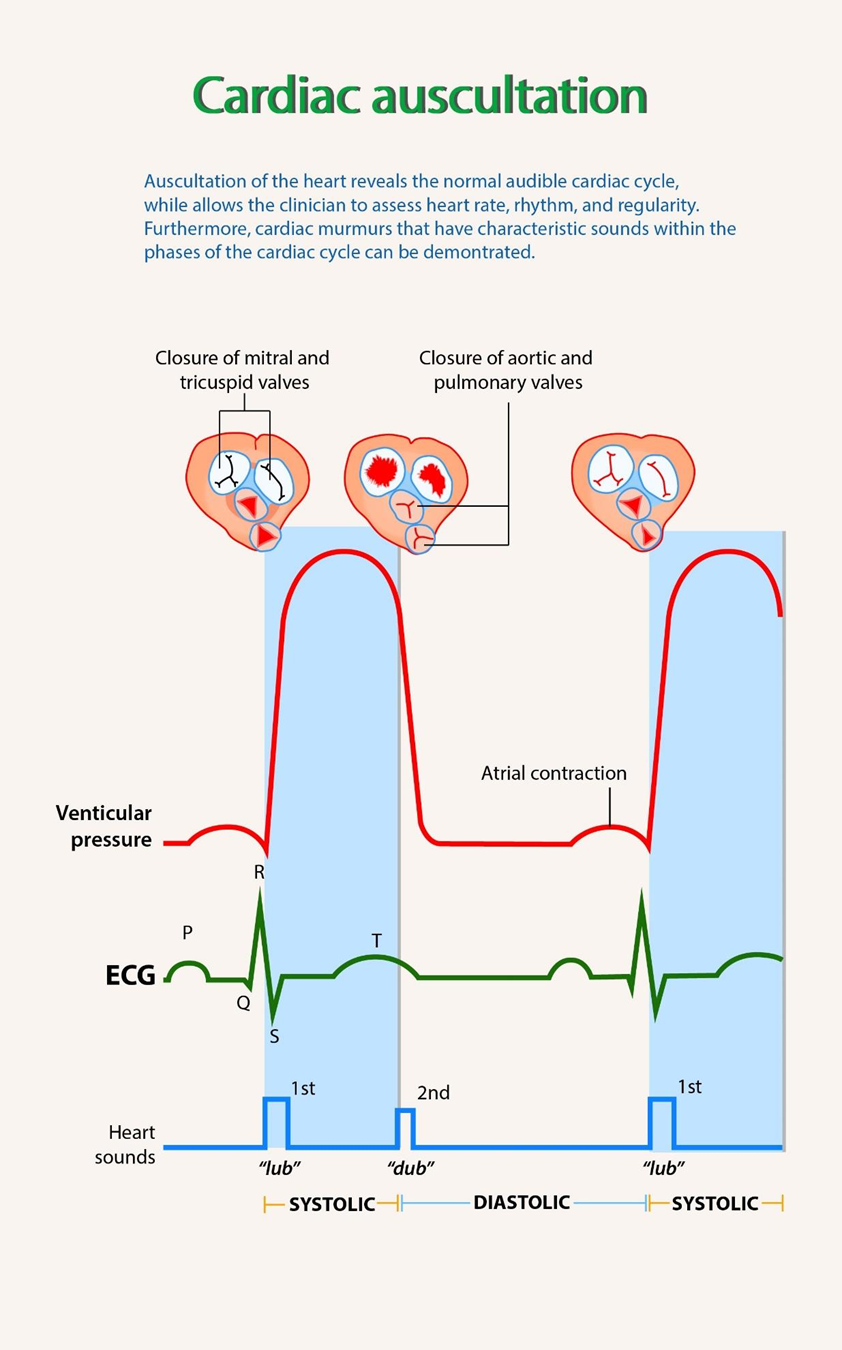
Choice A reason: The S1 heart sound represents the closure of the atrioventricular valves, and S2 represents the closure of the semilunar valves. The interval between S1 and S2 corresponds to ventricular systole. Any audible turbulence or vibration detected during this specific cardiac phase is classified as a systolic murmur, often resulting from increased blood flow or valvular pathology.
Choice B reason: While S1 and S2 are considered expected heart sounds, turbulence audible during the systolic phase is considered an adventitious sound. An expected or normal heart examination typically involves clear, crisp valvular closures without audible swishing or blowing sounds, which indicate abnormal blood flow patterns.
Choice C reason: The third heart sound is a low-frequency sound occurring during the rapid filling phase of early diastole, immediately following S2. It is produced by the vibration of the ventricular walls as blood rushes into the ventricles. It is not associated with the systolic interval between S1 and S2.
Choice D reason: The fourth heart sound is a low-pitched sound heard in late diastole, just before S1. It is generated by the atrial contraction forcing blood into a stiff, non-compliant ventricle. Like the S3, this sound occurs during diastole and is not related to the turbulence occurring during the systolic interval.
Whether you are a student looking to ace your exams or a practicing nurse seeking to enhance your expertise , our nursing education contents will empower you with the confidence and competence to make a difference in the lives of patients and become a respected leader in the healthcare field.
Visit Naxlex, invest in your future and unlock endless possibilities with our unparalleled nursing education contents today