Which structure is highlighted blue and indicated by the arrows in the image below?
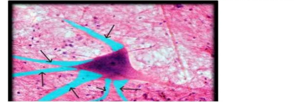
Axon terminal
Axon
Dendrite
Cell body
The Correct Answer is C
The marked structure is the dendrite, a branched projection of a neuron that extends from the cell body (soma). Dendrites are specialized for receiving synaptic input from other neurons and transmitting that electrical signal toward the cell body. They increase the surface area available for synaptic connections, allowing integration of multiple incoming signals. Functionally, dendrites play a key role in determining whether a neuron reaches the threshold for action potential generation at the axon hillock.
A. Axon terminal: The axon terminal is the distal end of the axon where neurotransmitters are released into the synaptic cleft. It forms synapses with other neurons, muscles, or glands to transmit signals to the next cell. Unlike dendrites, which receive input, axon terminals are involved in signal output. They are usually found at the far end of long axonal projections rather than branching near the cell body.
B. Axon: The axon is a long, single projection that carries electrical impulses away from the neuron’s cell body toward target cells. It is often myelinated to increase conduction speed and ends in terminal branches. Unlike dendrites, it is typically singular, longer, and specialized for signal transmission rather than reception.
C. Dendrite: Dendrites are multiple, short, highly branched extensions of the neuron that receive incoming synaptic signals. They conduct graded potentials toward the soma, where integration occurs to determine neuronal firing. Their extensive branching increases receptive surface area, making them essential for neural communication. Their structure and function as primary input receivers make them the correct answer.
D. Cell body: The cell body (soma) contains the nucleus and most organelles required for neuronal metabolism and protein synthesis. It integrates incoming signals from dendrites and maintains cell function. Unlike dendrites, it is not a branching structure but a central region of the neuron. It serves as the metabolic center rather than the primary input surface.
Nursing Test Bank
Naxlex Comprehensive Predictor Exams
Related Questions
Correct Answer is C
Explanation
The marked structure is the deltoid muscle, a large, thick, triangular muscle covering the shoulder joint and forming the rounded contour of the shoulder. It originates from the lateral third of the clavicle, the acromion, and the spine of the scapula, and inserts on the deltoid tuberosity of the humerus. The deltoid is the primary abductor of the arm at the glenohumeral joint, especially beyond the initial 15 degrees initiated by the supraspinatus. It is also involved in flexion, extension, and rotation of the shoulder depending on the muscle fibers activated.
A. Trapezius: The trapezius is a large, superficial muscle of the upper back extending from the occipital bone to the lower thoracic vertebrae and laterally to the scapula and clavicle. It functions in scapular elevation, retraction, depression, and rotation, contributing to posture and shoulder stabilization. Unlike the deltoid, it does not act directly on the humerus or produce shoulder abduction.
B. Biceps brachii: The biceps brachii is located in the anterior compartment of the upper arm and has two heads originating from the scapula. It primarily functions in elbow flexion and forearm supination. It is not a shoulder muscle and does not form the rounded contour of the shoulder like the deltoid.
C. Deltoid: The deltoid is a multipennate muscle covering the lateral shoulder, forming its rounded contour. It abducts the arm at the shoulder joint and assists in flexion, extension, and rotation depending on fiber orientation. It originates from the clavicle, acromion, and scapular spine and inserts on the humerus. Its superficial position and shoulder-covering shape make it the correct identification.
D. Latissimus dorsi: The latissimus dorsi is a broad, flat muscle of the back that extends from the lower thoracic spine, lumbar fascia, and iliac crest to the humerus. It functions in shoulder extension, adduction, and internal rotation. Compared to the deltoid, it is located posteriorly and inferiorly and does not form the shoulder’s rounded contour.
Correct Answer is A
Explanation
The skin is composed of three main layers: the epidermis, dermis, and hypodermis (subcutaneous layer). The epidermis is the outermost layer and is responsible for forming a protective barrier against environmental damage. It undergoes continuous renewal through a process called keratinization, where cells move from the basal layer to the surface and are eventually shed. Disorders affecting desquamation, such as ichthyosis, primarily involve abnormalities in this outer epithelial layer, leading to accumulation of dead keratinized cells and scaly skin.
A. Epidermis: ichthyosis affects the epidermis, specifically the stratum corneum, which is the outermost portion of the epidermis. Normally, keratinocytes undergo a regulated process of differentiation and desquamation, where dead cells are shed from the skin surface. In ichthyosis, this process is disrupted, leading to excessive accumulation of keratinized cells and a thick, scaly appearance. The epidermis is responsible for barrier function and continuous renewal, making it the primary site of pathology.
B. Dermis: The dermis is the deeper layer of the skin located beneath the epidermis and is composed of connective tissue containing collagen, elastin, blood vessels, nerves, and hair follicles. It provides structural support, elasticity, and nourishment to the epidermis. While it plays an important supportive role, it is not involved in keratinization or surface cell shedding.
C. Hypodermis: The hypodermis, also known as the subcutaneous layer, is the deepest layer of the skin and is primarily composed of adipose tissue and loose connective tissue. It functions in insulation, energy storage, and cushioning of underlying structures. It does not participate in epidermal cell turnover or keratinization. As a result, it is not involved in the pathological process seen in ichthyosis.
D. Subcutaneous layer: The subcutaneous layer is another term for the hypodermis and shares the same structure and functions. It lies beneath the dermis and consists mainly of fat and connective tissue. Its role is primarily supportive and metabolic rather than epithelial renewal. Since ichthyosis is a disorder of epidermal desquamation, the subcutaneous layer is not involved in this condition.
Whether you are a student looking to ace your exams or a practicing nurse seeking to enhance your expertise , our nursing education contents will empower you with the confidence and competence to make a difference in the lives of patients and become a respected leader in the healthcare field.
Visit Naxlex, invest in your future and unlock endless possibilities with our unparalleled nursing education contents today