
ANATOMY and PHYSIOLOGY. Skeletal Tissue, The Skeleton, Joints Proctored Exam
Total Questions : 44
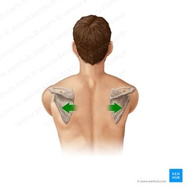
Showing 10 questions, Sign in for moreIdentify the anatomical movement illustrated below and state the joint involved (for example flexion of the knee joint).
Explanation
Scapula protraction refers to the movement of the shoulder blade (scapula) away from the spine, toward the front of the body.
This movement is also known as scapular abduction or anterior scapular tilt.
The joint involved in scapula protraction is the scapulothoracic joint, which is not a true joint, but rather a functional joint formed by the articulation between the scapula and the thorax.
The scapula is a flat bone that glides over the back of the ribcage, allowing for a wide range of movements of the arm.
Other movements of the scapulothoracic joint include:
Scapula retraction: This refers to the movement of the shoulder blade towards the spine, away from the front of the body.
This movement is also known as scapular adduction or posterior scapular tilt.
Scapula elevation: This refers to the movement of the shoulder blade upwards towards the ears.
This movement is also known as the upward rotation of the scapula.
Scapula depression: This refers to the movement of the shoulder blade downwards towards the feet.
This movement is also known as a downward rotation of the scapula.
Scapula upward tilt: This refers to the movement of the upper border of the shoulder blade upwards, towards the head.
This movement is also known as superior scapular rotation.
Scapula downward tilt: This refers to the movement of the upper border of the shoulder blade downwards, towards the feet.
This movement is also known as inferior scapular rotation.
All of these movements are important for proper shoulder function and are necessary for a wide range of daily activities, such as reaching, lifting, pushing, and pulling.
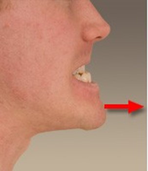
Identify the anatomical movement illustrated below and state the joint involved (for example flexion of the knee joint).
Explanation
Mandible protraction refers to the movement of the lower jaw (mandible) forwards, away from its rest position.
This movement is also known as jaw thrust.
The joint involved in mandible protraction is the temporomandibular joint (TMJ), which is a synovial joint that connects the mandible to the temporal bone of the skull.
This joint allows for a variety of movements, including:
Mandible retraction: This refers to the movement of the lower jaw backward, towards the skull.
Mandible elevation: This refers to the movement of the lower jaw upwards, towards the upper jaw.
Mandible depression: This refers to the movement of the lower jaw downwards, away from the upper jaw.
Mandible lateral excursion: This refers to the movement of the lower jaw to either the left or the right.
Mandible medial excursion: This refers to the movement of the lower jaw back to its midline position after a lateral excursion.
These movements of the TMJ are essential for functions such as chewing, speaking, and swallowing.
However, excessive or repetitive movements of the jaw can lead to TMJ disorders, which can cause pain, clicking, popping, or locking of the jaw.
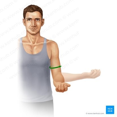
Identify the anatomical movement illustrated below and state the joint involved (for example flexion of the knee joint).

Explanation
Radial flexion of the wrist, also known as radial deviation, refers to the movement of the wrist towards the radial (thumb) side of the forearm.
This movement is achieved by the contraction of muscles on the lateral (thumb) side of the forearm.
The joint involved in radial flexion of the wrist is the radiocarpal joint, which is a condyloid joint formed by the articulation between the distal end of the radius bone and the carpal bones of the wrist.
This joint allows for a range of movements, including:
1. Ulnar flexion of the wrist: This refers to the movement of the wrist towards the ulnar (little finger) side of the forearm, which is achieved by the contraction of muscles on the medial (little finger) side of the forearm.
2. Extension of the wrist: This refers to the movement of the wrist backward, away from the palm of the hand.
This movement is achieved by the contraction of muscles on the back of the forearm.
3. Flexion of the wrist: This refers to the movement of the wrist forwards, towards the palm of the hand.
This movement is achieved by the contraction of muscles on the front of the forearm.
4. Adduction of the wrist: This refers to the movement of the wrist towards the midline of the body, which is achieved by the contraction of muscles on the medial (little finger) side of the forearm.
5. Abduction of the wrist: This refers to the movement of the wrist away from the midline of the body, towards the radial (thumb) side of the forearm, which is achieved by the contraction of muscles on the lateral (thumb) side of the forearm.
All of these movements of the radiocarpal joint are important for fine motor skills, such as writing, typing, and playing musical instruments, as well as for everyday activities such as lifting and carrying objects.
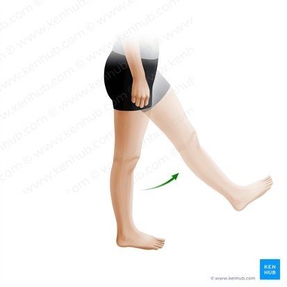
Identify the anatomical movement illustrated below and state the joint involved (for example flexion of the knee joint).
Explanation
Hip flexion refers to the movement of the thigh bone (femur) towards the front of the body, bringing the knee closer to the chest.
This movement is achieved by the contraction of muscles in the front of the hip, including the iliopsoas and rectus femoris muscles.
The joint involved in hip flexion is the hip joint, which is a ball-and-socket joint formed by the articulation between the head of the femur and the acetabulum of the pelvis.
This joint allows for a range of movements, including:
1. Hip extension: This refers to the movement of the thigh bone backwards, away from the front of the body, which is achieved by the contraction of muscles in the back of the hip, including the gluteus maximus and hamstrings.
2. Hip abduction: This refers to the movement of the thigh bone away from the midline of the body, towards the side, which is achieved by the contraction of muscles on the outside of the hip, including the gluteus medius and tensor fasciae latae.
3. Hip adduction: This refers to the movement of the thigh bone towards the midline of the body, which is achieved by the contraction of muscles on the inside of the hip, including the adductor muscles.
4. Hip external rotation: This refers to the movement of the thigh bone away from the midline of the body and outwards, which is achieved by the contraction of muscles on the back of the hip, including the piriformis and gemellus muscles.
5. Hip internal rotation: This refers to the movement of the thigh bone towards the midline of the body and inwards, which is achieved by the contraction of muscles on the inside of the hip, including the gluteus minimus and tensor fasciae latae.
All of these movements of the hip joint are important for walking, running, and other activities that require the use of the lower limbs.
Additionally, a lack of hip flexibility or strength can lead to compensatory movements in other parts of the body, which can result in pain or injury over time.
Identify the anatomical movement illustrated below and state the joint involved (for example flexion of the knee joint).
Explanation
External shoulder rotation is the movement of the humerus bone away from the centerline of the body, rotating the arm so that the palm faces away from the body.
This movement is achieved by the contraction of the external rotator muscles of the shoulder, which include the infraspinatus, teres minor, and posterior fibers of the deltoid muscle.
The joint involved in external shoulder rotation is the glenohumeral joint, also known as the shoulder joint.
The shoulder joint is a ball-and-socket joint formed by the articulation between the head of the humerus bone and the glenoid cavity of the scapula bone.
This joint allows for a range of movements, including:
1. Internal shoulder rotation: This is the opposite movement of external rotation, in which the humerus bone rotates inward toward the centerline of the body, with the palm facing inward.
2. Shoulder flexion: This is the movement of the humerus bone forward and upward, bringing the arm closer to the body's front.
3. Shoulder extension: This is the movement of the humerus bone backward and downward, moving the arm away from the body's front.
4. Shoulder abduction: This is the movement of the humerus bone away from the body's midline, raising the arm out to the side.
5. Shoulder adduction: This is the movement of the humerus bone toward the body's midline, bringing the arm back down to the side of the body.
6. Shoulder horizontal abduction: This is the movement of the humerus bone away from the body's midline at shoulder height.
7. Shoulder horizontal adduction: This is the movement of the humerus bone toward the body's midline at shoulder height.
All of these movements of the glenohumeral joint are important for many daily activities, including reaching, throwing, pushing, pulling, and lifting.
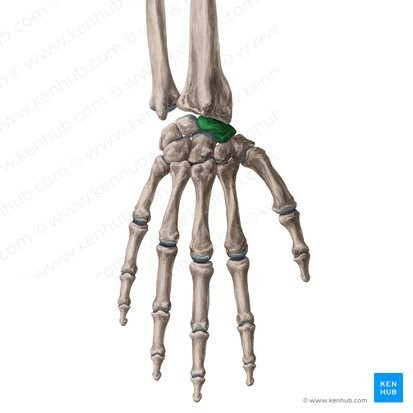
Identify the bone in the diagram below
Explanation
The carpal bones, also known as the wrist bones, are a group of eight small bones located in the wrist joint.
They are arranged in two rows of four bones each, with the rows separated by a space known as the carpal tunnel.
The carpal bones are held together by ligaments, and their shape and arrangement allow for a wide range of wrist movements.
The names of the carpal bones, from the proximal row to the distal row, are the scaphoid, lunate, triquetrum, and pisiform, and the trapezium, trapezoid, capitate, and hamate.
Each bone has a unique shape and surface features that allow it to articulate with adjacent bones, forming a complex network of joints that are important for wrist and hand movements.
The carpal bones are important because they provide stability to the wrist joint, allowing for precise movements of the hand and fingers.
They also help to transfer forces from the hand to the forearm, and vice versa.
Injuries to the carpal bones can result in wrist pain, instability, and decreased function of the hand and fingers.
Additionally, the arrangement of the carpal bones can affect the function of the median nerve, which runs through the carpal tunnel.
Compression or irritation of this nerve can result in carpal tunnel syndrome, a condition characterized by pain, numbness, and tingling in the hand and fingers.
Identify the bone in the diagram below
Explanation
The tibia, also known as the shinbone, is the larger of the two bones in the lower leg.
It is a long bone that extends from the knee joint to the ankle joint, and it plays an important role in supporting body weight and providing attachment sites for muscles.
The tibia has a triangular cross-section and features several surface landmarks, including the tibial plateau, the medial malleolus, and the tibial tuberosity.
The bone is also involved in the formation of the knee joint and the ankle joint, where it articulates with the femur and the talus bones, respectively.
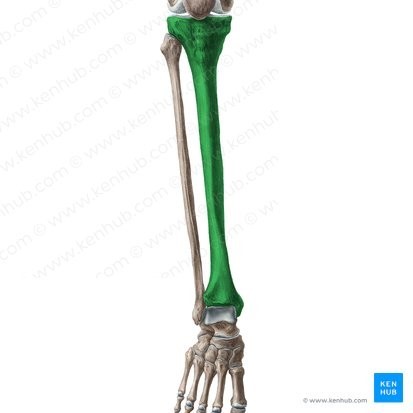
Identify the bone in the diagram below
Explanation

The occipital bone is a flat, unpaired bone located at the posterior aspect of the skull, forming the lower part of the back of the head.
It features several surface landmarks, including the external occipital protuberance, which serves as an attachment site for muscles and ligaments.
The occipital bone also contains several foramina, including the foramen magnum, which allows the spinal cord to pass through and connect to the brain.
The occipital bone is an important site for the attachment of muscles involved in head movement and posture.
Identify the structure in the diagram below
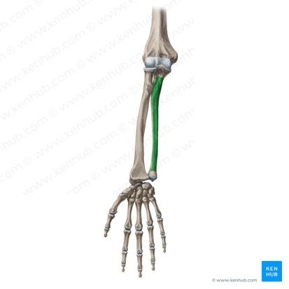
Explanation
The ulna is one of the two bones of the forearm, located on the medial (inner) side of the arm.
It is a long bone that runs parallel to the radius bone, extending from the elbow joint to the wrist joint.
The ulna features several surface landmarks, including the olecranon process, which forms the bony tip of the elbow.
The bone is also involved in the formation of the elbow joint, where it articulates with the humerus bone, and the wrist joint, where it articulates with the radius bone and several carpal bones.
The ulna is an important site for the attachment of muscles involved in forearm and wrist movements.
Identify the structure in the diagram below

Explanation
The posterior sacral foramina are openings located on the posterior aspect of the sacrum bone, which is part of the pelvis.
They are paired structures that allow the sacral nerves and blood vessels to pass through and exit the sacrum.
There are typically four pairs of posterior sacral foramina, located at the lateral margins of the sacrum.
These foramina are important for the transmission of nerve signals and blood supply to the lower limbs and pelvic organs.
Sign Up or Login to view all the 44 Questions on this Exam
Join over 100,000+ nursing students using Naxlex’s science-backend flashcards, practice tests and expert solutions to improve their grades and reach their goals.
Sign Up Now
