A nurse is collecting data from an infant who has coarctation of the aorta. Which of the following manifestations should the nurse expect?
Machine-like murmur
Severe cyanosis
Decreased blood pressure in the legs
Pulmonary edema
The Correct Answer is C
A. Machine-like murmur:
A machine-like murmur typically refers to a continuous murmur, which can be heard throughout systole and diastole. While machine-like murmurs can be associated with certain cardiac conditions, such as patent ductus arteriosus (PDA), they are not typically heard in coarctation of the aorta. In coarctation of the aorta, a systolic ejection murmur may be heard over the upper left sternal border due to turbulent blood flow across the narrowed aortic segment.
B. Severe cyanosis:
Cyanosis refers to a bluish discoloration of the skin and mucous membranes due to decreased oxygenation of the blood. While cyanosis can occur in various congenital heart defects, such as tetralogy of Fallot, it is not a characteristic manifestation of coarctation of the aorta. Coarctation of the aorta typically results in decreased blood flow to the lower extremities rather than mixing of oxygenated and deoxygenated blood.
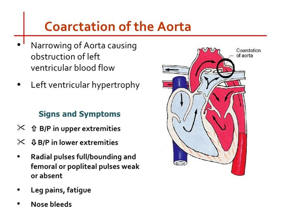
C. Decreased blood pressure in the legs:
This is the correct choice. Coarctation of the aorta is characterized by narrowing of the aorta, which leads to decreased blood flow to the lower extremities. Consequently, blood pressure measurements in the legs may be lower compared to those in the arms. This finding is often a key indicator of coarctation of the aorta.
D. Pulmonary edema:
Pulmonary edema refers to the accumulation of fluid in the lungs and is typically associated with conditions such as heart failure or fluid overload. While some congenital heart defects may lead to heart failure and subsequent pulmonary edema, coarctation of the aorta does not directly cause pulmonary edema. Instead, it primarily affects blood flow to the lower extremities due to the narrowing of the aorta.
Nursing Test Bank
Naxlex Comprehensive Predictor Exams
Related Questions
Correct Answer is A
Explanation
A. Give the child a stuffed animal: Providing a comforting item like a stuffed animal can help the child feel more secure and may offer some comfort during the parent's absence. This option is appropriate as it addresses the child's emotional needs.
B. Inform the child that her parent will be back in 2 hours: While it's helpful to provide reassurance to the child, a 2-year-old may not fully understand the concept of time, and telling them that their parent will return in 2 hours may not effectively alleviate their distress. This option may not be as immediately comforting as providing a tangible source of comfort.
C. Call the parent to return to the child's room: If possible, having the parent return to the child's room can provide the most direct comfort and reassurance to the child during a tantrum. However, it may not always be feasible for the parent to return immediately, especially if they are occupied or attending to other responsibilities.
D. Leave the child alone in the room for 5 minutes: Leaving the child alone during a tantrum can exacerbate feelings of distress and abandonment, potentially escalating the situation further. It's essential to provide support and reassurance to the child during moments of distress rather than leaving them alone.
Correct Answer is C
Explanation
A. Administer the medication at mealtime.Ferrous sulfate is best absorbed on an empty stomach because food, especially those rich in calcium or tannins, can interfere with its absorption. Administering it with meals reduces its effectiveness.
B.While bedtime administration is not contraindicated, it is not necessary. The timing of administration should focus on maximizing absorption, typically between meals or on an empty stomach.
C. Ferrous sulfate can stain teeth if taken orally in liquid form. Using a straw minimizes contact with teeth, reducing the risk of discoloration. Parents should also be advised to encourage the child to rinse their mouth after taking the medication.
D. Dilute the medication with 240 mL of milk. Milk contains calcium, which inhibits the absorption of iron. Ferrous sulfate should not be taken with milk or dairy products to ensure optimal absorption.

Whether you are a student looking to ace your exams or a practicing nurse seeking to enhance your expertise , our nursing education contents will empower you with the confidence and competence to make a difference in the lives of patients and become a respected leader in the healthcare field.
Visit Naxlex, invest in your future and unlock endless possibilities with our unparalleled nursing education contents today