Identify the bone in the diagram below
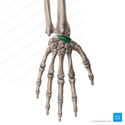
The Correct Answer is ["Scaphoid bone"]
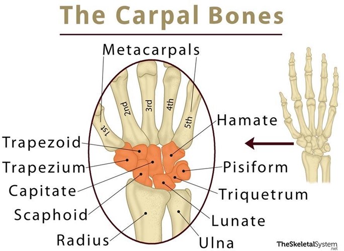
The carpal bones, also known as the wrist bones, are a group of eight small bones located in the wrist joint.
They are arranged in two rows of four bones each, with the rows separated by a space known as the carpal tunnel.
The carpal bones are held together by ligaments, and their shape and arrangement allow for a wide range of wrist movements.
The names of the carpal bones, from the proximal row to the distal row, are the scaphoid, lunate, triquetrum, and pisiform, and the trapezium, trapezoid, capitate, and hamate.
Each bone has a unique shape and surface features that allow it to articulate with adjacent bones, forming a complex network of joints that are important for wrist and hand movements.
The carpal bones are important because they provide stability to the wrist joint, allowing for precise movements of the hand and fingers.
They also help to transfer forces from the hand to the forearm, and vice versa.
Injuries to the carpal bones can result in wrist pain, instability, and decreased function of the hand and fingers.
Additionally, the arrangement of the carpal bones can affect the function of the median nerve, which runs through the carpal tunnel.
Compression or irritation of this nerve can result in carpal tunnel syndrome, a condition characterized by pain, numbness, and tingling in the hand and fingers.
Nursing Test Bank
Naxlex Comprehensive Predictor Exams
Related Questions
Correct Answer is B
Explanation
The ethmoid bone is a part of the axial skeleton, not the appendicular skeleton.
The axial skeleton consists of the bones of the skull, vertebral column, ribs, and sternum.
Choice A is incorrect because the ulna is a part of the upper limb, which is supported by the pectoral girdle.
Choice C is incorrect because the ilium is a part of the hip bone, which forms the pelvic girdle.
Choice D is incorrect because the patella is a part of the lower limb, which is supported by the pelvic girdle.
Correct Answer is A
Explanation
The cervical vertebrae are the only vertebrae that have transverse foramina, which are openings in the transverse processes that allow the passage of the vertebral arteries and veins.
Choice B is incorrect because lumbar vertebrae do not have transverse foramina.
They have large bodies and short, thick transverse processes that serve as attachment sites for muscles.
Choice C is incorrect because thoracic vertebrae do not have transverse foramina.
They have costal facets on their transverse processes that articulate with the tubercles of the ribs.
Choice D is incorrect because sacral vertebrae do not have transverse foramina.
They are fused together to form the sacrum, which has four pairs of sacral foramina on each side that transmit sacral nerves and vessels.
Whether you are a student looking to ace your exams or a practicing nurse seeking to enhance your expertise , our nursing education contents will empower you with the confidence and competence to make a difference in the lives of patients and become a respected leader in the healthcare field.
Visit Naxlex, invest in your future and unlock endless possibilities with our unparalleled nursing education contents today