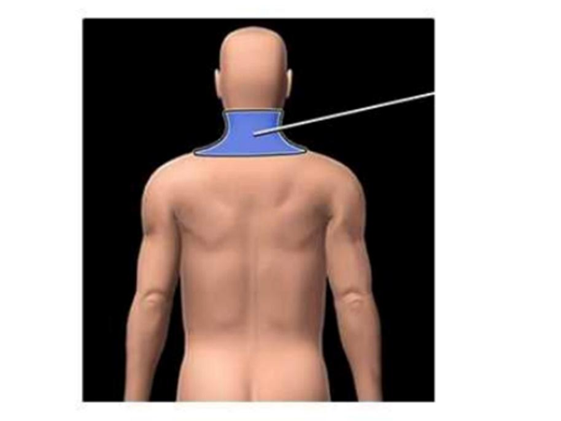
Identify the highlighted region.
Acromial region
Cervical region
Vertebral region
Gluteal region
Lumbar region
The Correct Answer is B
The marked structure is the cervical region, which corresponds anatomically to the neck portion of the vertebral column and surrounding soft tissues. It extends from the base of the skull to the level of the first thoracic vertebra (C1–C7). This region supports the head, allows a wide range of head and neck movements, and provides a passage for critical structures including the trachea, esophagus, carotid arteries, jugular veins, and cervical spinal cord. It also contains important muscle groups such as the sternocleidomastoid and scalene muscles that contribute to posture, respiration, and head mobility.
A. Acromial region: The acromial region refers to the lateral aspect of the shoulder over the acromion process of the scapula. It forms part of the shoulder girdle and serves as an attachment point for the deltoid muscle. Unlike the cervical region, it is located on the upper lateral shoulder rather than the neck and is primarily involved in upper limb movement.
B. Cervical region: The cervical region is the neck portion of the body consisting of the cervical vertebrae and surrounding soft tissues. It supports the head, allows flexion, extension, rotation, and lateral bending, and houses vital neurovascular and airway structures. It forms the transition between the skull and thorax, making it essential for both structural support and communication pathways. Its location corresponds directly with the highlighted neck area.
C. Vertebral region: The vertebral region refers broadly to the entire spinal column, including cervical, thoracic, lumbar, sacral, and coccygeal segments. It provides axial support and protection for the spinal cord. Unlike the cervical region, it is not limited to the neck but spans the entire back from skull to pelvis.
D. Gluteal region: The gluteal region refers to the buttock area, composed mainly of the gluteus maximus, medius, and minimus muscles. It plays a key role in hip movement and locomotion. Compared to the cervical region, it is located in the lower posterior trunk and is unrelated to neck structures.
E. Lumbar region: The lumbar region refers to the lower back area containing the lumbar vertebrae (L1–L5). It supports body weight and allows trunk flexion and extension. Unlike the cervical region, it is situated between the thoracic spine and sacrum, forming the lower posterior trunk rather than the neck.
F. Sacral region: The sacral region is located at the base of the spine and consists of the fused sacral vertebrae forming the sacrum. It contributes to pelvic stability and transfers weight from the spine to the lower limbs. Compared to the cervical region, it is positioned inferiorly within the pelvis rather than in the neck.
Nursing Test Bank
Naxlex Comprehensive Predictor Exams
Related Questions
Correct Answer is B
Explanation
Skeletal muscle contraction depends on excitation–contraction coupling, a process where an electrical signal triggers calcium release, leading to interaction between actin and myosin filaments. Calcium ions are essential because they bind to troponin, shifting tropomyosin away from actin binding sites and allowing cross-bridge formation. The rapid availability of calcium within muscle fibers is ensured by specialized intracellular storage systems. The sarcoplasmic reticulum plays a central role in regulating calcium release and reuptake to control contraction and relaxation.
A. Mitochondria: Mitochondria are organelles responsible for ATP production through aerobic respiration, specifically oxidative phosphorylation. While they are essential for providing energy required for muscle contraction, they do not serve as a primary calcium storage or release site for contraction initiation. Although mitochondria can take up calcium for metabolic regulation, this is not the calcium source that activates troponin.
B. Sarcoplasmic reticulum: the sarcoplasmic reticulum (SR) is the specialized intracellular organelle that stores and releases calcium ions in skeletal muscle fibers. When an action potential travels along the sarcolemma and into the T-tubules, it triggers voltage-sensitive receptors that open calcium channels in the SR. Calcium then floods into the sarcoplasm and binds to troponin C, initiating the contraction process. The SR acts as the primary and rapid regulatory calcium reservoir for muscle excitation–contraction coupling.
C. Extracellular fluid: Extracellular fluid contains calcium ions, but skeletal muscle contraction does not depend on extracellular calcium influx as the primary trigger. Unlike cardiac muscle, skeletal muscle relies mainly on internal calcium stores from the sarcoplasmic reticulum. The small amount of extracellular calcium present is not sufficient or directly responsible for initiating contraction. This source is not the primary contributor to troponin activation in skeletal muscle.
D. T-tubules: T-tubules are invaginations of the sarcolemma that transmit the action potential deep into the muscle fiber. Their role is electrical conduction rather than calcium storage or release. They are structurally linked to the sarcoplasmic reticulum and help trigger calcium release via voltage-sensitive proteins. However, they do not contain or supply calcium themselves, making them an incorrect source of calcium ions for contraction.
Correct Answer is C
Explanation
Broca’s area is a specialized region of the dominant frontal lobe (usually the left hemisphere) located in the inferior frontal gyrus. It plays a critical role in the motor production of speech and language formulation. This area coordinates the complex motor planning required to articulate words, allowing fluent verbal expression. Damage to this region disrupts speech output while often preserving comprehension, resulting in a characteristic language deficit known as expressive aphasia.
A. Memory loss: memory processing is primarily associated with the hippocampus and medial temporal lobe structures. These areas are responsible for forming and retrieving long-term memories. Broca’s area does not play a direct role in memory storage or recall. Therefore, damage to Broca’s area would not primarily result in memory impairment.
B. Impaired motor control of the left side of the body: motor control of the body is governed by the primary motor cortex in the frontal lobe, particularly the precentral gyrus. Additionally, motor pathways decussate in the brainstem, meaning the right hemisphere controls the left side of the body. While nearby motor regions may be involved in movement, Broca’s area is specifically dedicated to speech production, not general motor control.
C. Inability to produce speech (expressive aphasia): Broca’s area is responsible for the motor planning and production of speech. Damage to this region results in expressive aphasia, where individuals understand language but struggle to form coherent spoken or written speech. Speech becomes slow, effortful, and fragmented, although comprehension remains relatively intact. This demonstrates the specialized role of Broca’s area in language expression.
D. Loss of visual processing: visual processing occurs in the occipital lobe, specifically the primary visual cortex. This region interprets visual stimuli received from the retina via the optic pathways. Broca’s area has no involvement in visual perception or interpretation. Therefore, damage to this area would not affect vision.
Whether you are a student looking to ace your exams or a practicing nurse seeking to enhance your expertise , our nursing education contents will empower you with the confidence and competence to make a difference in the lives of patients and become a respected leader in the healthcare field.
Visit Naxlex, invest in your future and unlock endless possibilities with our unparalleled nursing education contents today