The largest artery in the body:
Carotid.
Aorta.
Celiac.
Femoral.
The Correct Answer is B
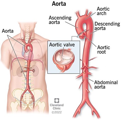
The aorta is the largest artery in the human body, as well as the main artery in the circulatory system.
It originates from the left ventricle of the heart and extends down to the abdomen, where it splits into two smaller arteries (the common iliac arteries).
The aorta distributes oxygenated blood to all parts of the body through the systemic circulation.
Choice A. Carotid is wrong because the carotid artery is not the largest artery in the body, but one of the main arteries that pumps blood from the heart to the brain and the rest of the head.
It has a diameter of 4.3 mm-7.7 mm and a blood flow of 350-550 milliliters per minute.
Choice C. Celiac is wrong because the celiac artery is not the largest artery in the body, but a major branch of the abdominal aorta that supplies oxygenated blood to the liver, stomach, spleen, pancreas, and duodenum.
Choice D. Femoral is wrong because the femoral artery is not the largest artery in the body, but the largest artery found in the leg region.
It runs down the inner thigh and carries out the important role of supplying blood to the lower body.
It has a diameter of 6.6 mm and a blood flow of 284 milliliters per minute.
Nursing Test Bank
Naxlex Comprehensive Predictor Exams
Related Questions
Correct Answer is D
Explanation
Exocrine glands secrete through ducts or tubes to the body’s exterior.
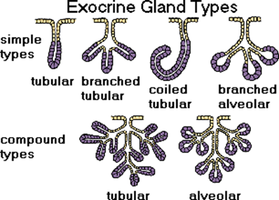
For example, sweat glands, salivary glands, and liver are exocrine glands.
Choice A is wrong because exocrine glands do not only secrete salts, but also other substances such as enzymes, mucus, and sebum.
Choice B is wrong because exocrine glands do not secrete hormones at all.
Hormones are secreted by endocrine glands, which are ductless glands that release their products directly into the bloodstream.
Choice C is wrong because exocrine glands do not secrete into the bloodstream, but onto an epithelial surface such as the skin or the gastrointestinal tract.
Only endocrine glands secrete into the bloodstream.
Correct Answer is B
Explanation
The correct answer is choice B. False.
Arteries are strong, elastic vessels that carry blood away from the heart, except for the coronary arteries that supply blood to the heart muscle.
These are the first arteries to branch off the aorta, which is the main artery that takes blood to the body from the left ventricle.
Choice A is wrong because it contradicts the definition of arteries. Arteries carry blood away from the heart, not to the heart.
Whether you are a student looking to ace your exams or a practicing nurse seeking to enhance your expertise , our nursing education contents will empower you with the confidence and competence to make a difference in the lives of patients and become a respected leader in the healthcare field.
Visit Naxlex, invest in your future and unlock endless possibilities with our unparalleled nursing education contents today