The nurse recognizes that the following statement is true regarding the internal structures of the breast: The breast is made up of:
Glandular tissue, which supports the breast by attaching to the chest wall.
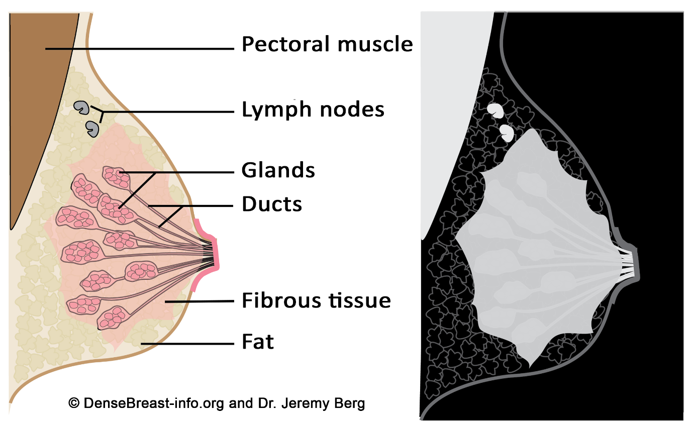
Fibrous, glandular, and adipose tissues
Primarily muscle with very little fibrous tissue.
Primarily milk ducts, known as lactiferous ducts.
The Correct Answer is B
A. Glandular tissue, which supports the breast by attaching to the chest wall: Glandular tissue is indeed a part of the breast structure, but it is not responsible for supporting the breast by attaching to the chest wall. It's the Cooper's ligaments, which are fibrous connective tissue, that provide structural support.
B. Fibrous, glandular, and adipose tissues: This statement is correct. The breast is composed of glandular tissue (responsible for milk production), fibrous tissue (including Cooper's ligaments for support), and adipose tissue (fat).
C. Primarily muscle with very little fibrous tissue: The breast contains very little muscle tissue. The main supportive structure is fibrous tissue, not muscle.
D. Primarily milk ducts, known as lactiferous ducts: Milk ducts are part of the glandular tissue and are responsible for carrying milk. However, the breast is not primarily made up of milk ducts; it consists of a combination of glandular, fibrous, and adipose tissues.
Nursing Test Bank
Naxlex Comprehensive Predictor Exams
Related Questions
Correct Answer is {"dropdown-group-1":"C"}
Explanation
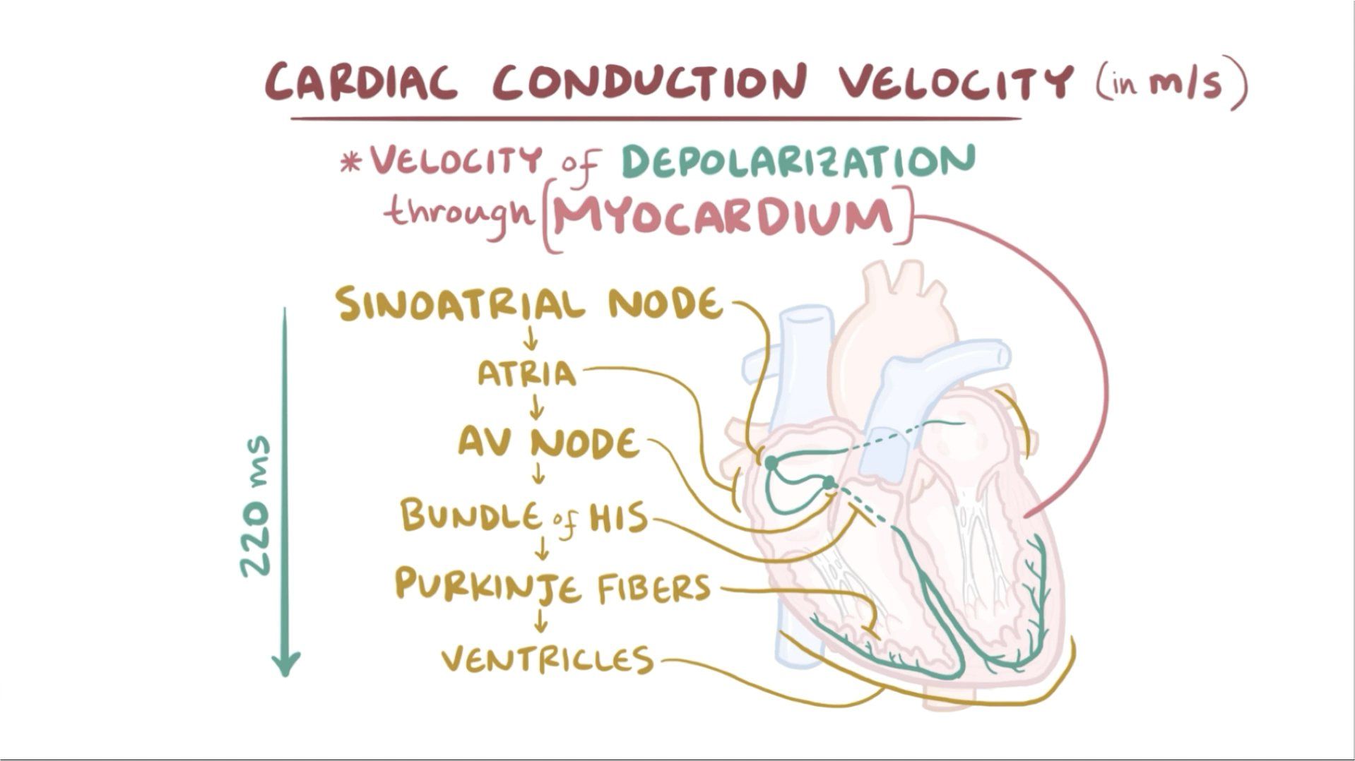
The pacemaker of the heart is known as the sinoatrial (SA) node. The SA node is a specialized group of cells located in the right atrium of the heart. It generates electrical impulses that initiate each heartbeat and set the rhythm for the entire heart.
Correct Answer is A
Explanation
A. A slight asymmetry in breast size can be expected: This response is accurate. It acknowledges the natural variation in breast size that many women experience. It's common for one breast to be slightly larger or shaped differently than the other. It assures the client that this asymmetry is normal and not a cause for concern.
B. Asymmetry of breast size and shape is probably due to breastfeeding and is nothing to worry about: While breastfeeding can cause temporary changes in breast size and shape, not all cases of breast asymmetry are related to breastfeeding. This statement might not cover all situations, making it less accurate.
C. A sudden uneven increase in breast size is normal in adults: This statement is not accurate. Sudden changes in breast size should always be investigated, as they can indicate underlying health issues and may not be considered normal.
D. Breasts should always be symmetric: This statement is not accurate. Perfect symmetry in breast size and shape is rare. Most women have some degree of asymmetry, which is entirely normal. It's important to reassure the client that slight differences are common and not a cause for concern.
Whether you are a student looking to ace your exams or a practicing nurse seeking to enhance your expertise , our nursing education contents will empower you with the confidence and competence to make a difference in the lives of patients and become a respected leader in the healthcare field.
Visit Naxlex, invest in your future and unlock endless possibilities with our unparalleled nursing education contents today