This is what type of tissue?
Simple Squamous Epithelium
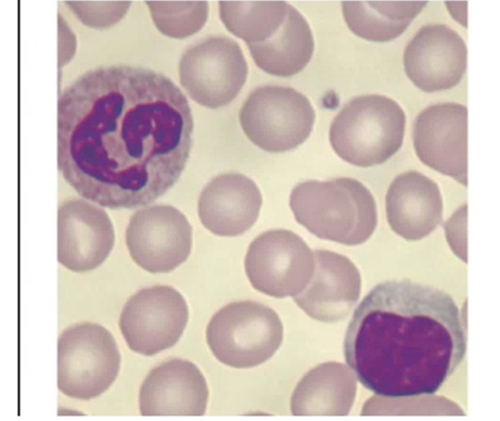
Connective Tissue (Blood)
Nervous Tissue
Smooth Muscle Tissue
The Correct Answer is B
A. Simple Squamous Epithelium: This tissue consists of a single layer of flat, scale-like cells that are tightly packed together to form a lining (like in the lungs or blood vessels). The cells in the image are free-floating and distinct from one another, which contradicts the "sheet-like" structure of epithelium.
B. Connective Tissue (Blood): Blood is classified as a specialized form of connective tissue because it consists of cells (formed elements) suspended in a non-living fluid matrix (plasma). Two larger cells with stained purple nuclei are visible-a neutrophil (top left with a multi-lobed nucleus) and a lymphocyte (bottom right with a large, round nucleus).
C. Nervous Tissue: Nervous tissue is characterized by neurons with long processes (axons and dendrites) and smaller supporting glial cells. The circular, isolated cells in this image do not possess the branching morphology typical of neural cells.
D. Smooth Muscle Tissue: Smooth muscle consists of elongated, spindle-shaped cells with a single central nucleus, usually arranged in tight sheets or bundles. The cells in the image are primarily rounded and dispersed, which is not characteristic of muscle fibers.
Nursing Test Bank
Naxlex Comprehensive Predictor Exams
Related Questions
Correct Answer is B
Explanation
A. Hair Follicles: These are the tube-like structures that house the hair root, seen extending deep into the dermis in the model, but they are not the "humps" at the interface.
B. Dermal Papillae: These are the finger-like projections of the dermis that indent the overlying epidermis. They increase surface area for nutrient exchange and form the basis for fingerprints.
C. Sebaceous Glands: These are oil-producing glands typically associated with hair follicles, visible as multi-lobed structures in the dermis, but they are not the "hump" structures indicated.
D. Hypodermal Ridges: The hypodermis is the deep fatty layer (yellow tissue at the bottom). Ridges are not a standard anatomical term for structures at that specific epidermal-dermal junction.
Correct Answer is D
Explanation
A. Simple squamous epithelium: Simple squamous epithelium consists of a single layer of flat cells, specialized for diffusion and filtration (e.g., alveoli, capillaries). It does not provide the protective layering needed in the vaginal canal.
B. Stratified squamous epithelium (non-keratinized): The vaginal canal is lined with non-keratinized stratified squamous epithelium. This multilayered tissue provides protection against friction and mechanical stress during intercourse and childbirth, while remaining moist and flexible.
C. Transitional epithelium: Transitional epithelium is found in the urinary bladder and ureters, where it allows stretching. It is not present in the vaginal canal.
D. Adipose tissue: The tissue shown at the arrow in the anatomical model is adipose tissue (commonly known as fat).The yellow, pebbled, or "honeycomb" texture is the standard representation of fat deposits on medical models.
Whether you are a student looking to ace your exams or a practicing nurse seeking to enhance your expertise , our nursing education contents will empower you with the confidence and competence to make a difference in the lives of patients and become a respected leader in the healthcare field.
Visit Naxlex, invest in your future and unlock endless possibilities with our unparalleled nursing education contents today