Identify the structure at the arrow.
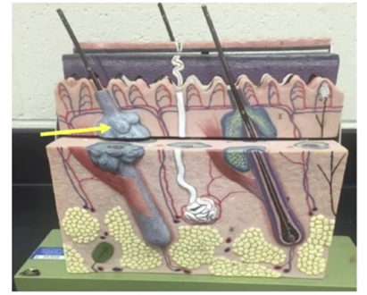
Sweat gland
Sebaceous gland
Hair follicle root
Arrector pili muscle
The Correct Answer is B
A. Sweat gland: Sweat glands are coiled tubular structures located deeper in the dermis or subcutaneous tissue. They do not appear as bulbous sacs attached directly to hair follicles.
B. Sebaceous gland: Sebaceous glands are sac-like structures that secrete sebum (oil) into the hair follicle. They are typically found adjacent to hair follicles, appearing as rounded, bulbous structures like the one indicated by the arrow.
C. Hair follicle root: The hair follicle root is the portion of the hair embedded in the dermis, extending downward into the follicular bulb. It is elongated, not bulbous, and is distinct from the glandular structure shown.
D. Arrector pili muscle: The arrector pili muscle is a thin band of smooth muscle attached to the hair follicle. It is not a glandular structure and does not have the rounded appearance of the sebaceous gland.
Nursing Test Bank
Naxlex Comprehensive Predictor Exams
Related Questions
Correct Answer is C
Explanation
- Sugar-phosphate backbone: This forms the vertical sides of the DNA ladder, not the horizontal rungs.
- Hydrogen ions: These are not structural components of DNA; hydrogen bonds hold base pairs together, but hydrogen ions themselves are not part of the DNA structure.
- Base pairs (nitrogenous base pairs): The horizontal rungs of the DNA ladder represent base pairs, formed by hydrogen bonding between complementary nitrogenous bases: adenine (A) pairs with thymine (T), and cytosine (C) pairs with guanine (G). These base pairs are critical for encoding genetic information and ensuring accurate replication. The arrow is not pointing to the entire DNA molecule, but specifically to the structural unit that connects the two strands
- Ribosomes: These are cellular organelles involved in protein synthesis, not part of DNA’s structure.
Correct Answer is A
Explanation
A. Stratum corneum: The stratum corneum is composed of dead, keratinized cells that form a tough, protective barrier. Its main function is to prevent water loss, protect against mechanical injury, and serve as the first line of defense against pathogens. Because it is the most superficial layer of the epidermis, it is the correct structure indicated by the arrow.
B. Stratum basale: This is the deepest layer of the epidermis, responsible for cell division and regeneration, not the outermost layer.
C. Dermal papillae: These are found in the dermis, not the epidermis. They interlock with the epidermis to strengthen the connection between the two layers.
D. Sebaceous gland: This gland is located in the dermis, associated with hair follicles, and secretes sebum. It is not part of the epidermis.
Whether you are a student looking to ace your exams or a practicing nurse seeking to enhance your expertise , our nursing education contents will empower you with the confidence and competence to make a difference in the lives of patients and become a respected leader in the healthcare field.
Visit Naxlex, invest in your future and unlock endless possibilities with our unparalleled nursing education contents today