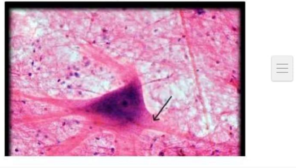
Which structure is indicated by the arrow in the image below?
Dendrite
Axon
Cell body (soma)
Synaptic terminal
The Correct Answer is B
The marked structure is the axon, a long, singular projection of a neuron that arises from the axon hillock of the cell body. It is specialized for conducting electrical impulses (action potentials) away from the soma toward target cells such as other neurons, muscle fibers, or glands. The axon may be myelinated, which increases conduction speed through saltatory conduction at the nodes of Ranvier. Its primary physiological role is rapid long-distance signal transmission within the nervous system.
A. Dendrite: Dendrites are short, highly branched projections that extend from the neuron cell body and function primarily to receive incoming synaptic signals. They transmit graded potentials toward the soma for integration. Unlike the axon, dendrites are typically multiple and tree-like in appearance, designed for input rather than output.
B. Axon: The axon is a single, elongated process that carries action potentials away from the neuron cell body toward synaptic targets. It is often myelinated to enhance conduction velocity, allowing rapid communication across long distances in the nervous system. It terminates in axon terminals that release neurotransmitters. Its structure as a long conducting fiber makes it the correct identification.
C. Cell body (soma): The cell body is the central part of the neuron that contains the nucleus and organelles responsible for metabolic and synthetic functions. It integrates signals received from dendrites and determines whether an action potential is initiated. Unlike the axon, it is not a projecting fiber but rather the central processing unit of the neuron.
D. Synaptic terminal: The synaptic terminal is the distal end of the axon where neurotransmitters are released into the synaptic cleft. It forms communication points with other neurons or effector cells. Unlike the axon, it is a terminal structure rather than the main conducting fiber and is responsible for signal transmission to the next cell rather than propagation along the neuron.
Nursing Test Bank
Naxlex Comprehensive Predictor Exams
Related Questions
Correct Answer is D
Explanation
Anatomical descriptions focus on the structure, location, and spatial relationships of body parts, while physiological descriptions explain how those structures function. In human biology, distinguishing between anatomy and physiology is essential for understanding how form relates to function. The heart and great vessels provide a good example, as they can be described either by their position and structure or by their functional properties such as contractility and endurance. Physiological statements specifically address what a structure does rather than where it is located.
A. The aorta is a large vessel connected to the heart: This is primarily an anatomical description because it identifies the aorta’s structure and its physical connection to the heart. It describes the size (large vessel) and its spatial relationship (attached to the heart), which are structural characteristics. It does not explain how the aorta functions in circulation, such as its role in carrying oxygenated blood under high pressure.
B. The heart is located in the thoracic cavity: This is strictly an anatomical description because it defines the location of the heart within the body. It specifies spatial positioning (thoracic cavity) without explaining any functional process. There is no reference to how the heart works, such as pumping blood or generating pressure.
C. The aorta lies anterior to the vertebral column in the thorax: This describes anatomical positioning using directional terms such as “anterior” and “vertebral column.” It explains the spatial relationship of the aorta to nearby structures. However, it does not describe any functional activity of the aorta, such as elastic recoil or blood flow regulation.
D. Heart muscle is under involuntary control and is fatigue resistant: This describes functional characteristics of cardiac muscle tissue. It explains how the heart operates automatically without conscious control and its ability to sustain continuous contractions without fatigue. These are physiological properties related to how the tissue functions rather than where it is located or how it is structured.
Correct Answer is A
Explanation
The vertebra is a complex irregular bone designed to provide structural support, flexibility, and protection for the spinal cord. It consists of a central body, a vertebral arch, and several specialized bony projections that serve as attachment points for muscles and ligaments. In the image, the structure marked with an "X" is one of the lateral projections extending from the vertebral arch, which is essential for stabilizing the vertebral column and facilitating movement through muscular pull.
A. The transverse process is the lateral bony projection that extends from the junction of the pedicle and lamina on each side of the vertebra. These processes serve as critical attachment sites for deep back muscles and, in the thoracic region, articulate with the ribs. The structure marked with an "X" is clearly identified as this lateral projection.
B. The spinous process is the singular, posterior-facing projection located at the midline of the vertebral arch. It is easily palpable through the skin along the back and serves as an anchor for various ligaments and muscles that support the spine. It is distinct from the lateral structure marked in the image, which originates from the side of the arch.
C. The superior articular facet is a smooth surface on the superior aspect of the vertebral arch that articulates with the inferior articular facet of the vertebra above it. These facets are crucial for limiting movement and maintaining the alignment of the vertebral column. They are located near the pedicle-lamina junction but are not the lateral extension identified by the "X" in the image.
D. The vertebral foramen is the large, central opening created by the vertebral arch and the posterior surface of the vertebral body. It houses the spinal cord, its associated protective meninges, and the surrounding blood vessels. The mark "X" is placed on the external lateral surface of the bone, not within this central canal.
Whether you are a student looking to ace your exams or a practicing nurse seeking to enhance your expertise , our nursing education contents will empower you with the confidence and competence to make a difference in the lives of patients and become a respected leader in the healthcare field.
Visit Naxlex, invest in your future and unlock endless possibilities with our unparalleled nursing education contents today