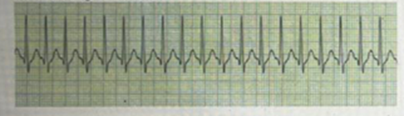
A client presents to the emergency department complaining of sudden onset of palpitations and chest pain. After assessment, the nurse notes the client to be diaphoretic, skin is cool to touch, BP is 80/40 mm Hg. Resp 26 and Sats of 89% [see image]. The nurse anticipates an order for which of the following interventions?
Syncronized cardioversion
Adenosine infusion over 30 minutes
Immediate defibrillation
Vagal manuevers
The Correct Answer is A
A. Synchronized cardioversion: Synchronized cardioversion is indicated for unstable supraventricular tachycardia (SVT), especially when the client shows signs of hemodynamic instability, such as hypotension, altered mental status, or chest pain. This intervention delivers a timed shock to restore normal rhythm, prioritizing the client's immediate stabilization.
B. Adenosine infusion over 30 minutes: Adenosine is typically administered as a rapid intravenous push to terminate SVT by temporarily blocking atrioventricular nodal conduction. However, this client is unstable, and synchronized cardioversion is the preferred intervention in cases of hemodynamic compromise.
C. Immediate defibrillation: Defibrillation is used for life-threatening arrhythmias like ventricular fibrillation or pulseless ventricular tachycardia. In this case, the rhythm is SVT, and the client is not in cardiac arrest, so defibrillation is inappropriate.
D. Vagal maneuvers: Vagal maneuvers, such as carotid sinus massage or the Valsalva maneuver, are first-line interventions for stable SVT. However, in unstable clients with severe symptoms or hemodynamic compromise, these measures are insufficient, and synchronized cardioversion is urgently required.
Nursing Test Bank
Naxlex Comprehensive Predictor Exams
Related Questions
Correct Answer is C
Explanation
A) A corticosteroid such as fluticasone:
While corticosteroids, such as fluticasone, are effective for managing chronic asthma and preventing inflammation over time, they are not the first-line treatment during an acute asthma attack. Corticosteroids are typically used for long-term control and maintenance therapy, not for rapid relief of symptoms in an acute exacerbation. Immediate relief is needed in acute situations, which corticosteroids alone do not provide.
B) A long-acting beta 2 agonist such as salmeterol:
Long-acting beta-2 agonists (LABAs), such as salmeterol, are used for maintenance therapy to prevent asthma attacks and should not be used for the immediate treatment of an acute asthma exacerbation. They take longer to start working, and their role is to provide prolonged bronchodilation over time, not to relieve sudden bronchoconstriction.
C) A short-acting beta 2 agonist such as albuterol:
During an acute asthma attack, the immediate goal is to relieve bronchoconstriction and improve airflow. Short-acting beta-2 agonists like albuterol are the first-line treatment because they quickly relax the smooth muscles of the airways, leading to bronchodilation. Albuterol works within minutes, providing rapid relief from the symptoms of wheezing, shortness of breath, and chest tightness.
D) Methylxanthines such as Theophylline:
Methylxanthines (e.g., theophylline) were once used for asthma management but are no longer considered the first-line treatment for acute exacerbations due to their narrow therapeutic range and the potential for toxicity. While theophylline can provide bronchodilation, its onset of action is slower than that of beta-agonists like albuterol, and it is generally reserved for more chronic management of asthma or severe cases where other medications are not effective.
Correct Answer is {"A":{"answers":"A"},"B":{"answers":"B"},"C":{"answers":"B"},"D":{"answers":"A"}}
Explanation
|
Assessment findings |
Expected Findings |
Findings to be reported to provider |
|
Barrel chest |
✔️ |
|
|
Increased fatigue |
✔️ |
|
|
Respiratory rate 40bpm |
✔️ |
|
|
Thin appearance |
✔️ |
Barrel chest: Expected Finding
Increased fatigue: Finding to be reported to the provider
Respiratory rate 40 bpm: Finding to be reported to the provider
Thin appearance: Expected Finding
Rationales:
Barrel chest – Expected Finding:
A barrel chest is a common physical finding in clients with chronic obstructive pulmonary disease (COPD), especially emphysema. It results from hyperinflation of the lungs over time, altering the shape of the chest wall.
Increased fatigue – Finding to be reported to the provider:
While COPD clients often experience fatigue, a sudden or unusual increase in fatigue may indicate worsening respiratory function or exacerbation of the disease. This finding requires further assessment and possible intervention to prevent complications.
Respiratory rate 40 bpm – Finding to be reported to the provider:
A respiratory rate of 40 bpm indicates significant tachypnea and respiratory distress. This finding, coupled with accessory muscle use, suggests the client may be experiencing an acute exacerbation of COPD or impending respiratory failure, which requires immediate provider notification.
Thin appearance – Expected Finding:
Clients with COPD often have a thin or cachectic appearance due to increased energy expenditure for breathing and reduced caloric intake. This is a typical finding in advanced COPD and does not require urgent reporting unless accompanied by other concerning symptoms.
Whether you are a student looking to ace your exams or a practicing nurse seeking to enhance your expertise , our nursing education contents will empower you with the confidence and competence to make a difference in the lives of patients and become a respected leader in the healthcare field.
Visit Naxlex, invest in your future and unlock endless possibilities with our unparalleled nursing education contents today