A woman is at 32 weeks' gestation. Her fundal height measurement at this clinic appointment is 26 centimeters. After reviewing her ultrasound results, the health care provider asks the nurse to schedule the client for a series of ultrasounds to be done every 2 weeks. The nurse should ensure that the client understands that the main purpose for this is to:
Select one:
Rule out a suspected hydatidiform mole.
Assess for congenital anomalies.
Determine fetal presentation.
Monitor fetal growth.
The Correct Answer is D
Choice A Reason: Rule out a suspected hydatidiform mole. This is an incorrect answer that describes an unlikely condition for this client. A hydatidiform mole is a type of gestational trophoblastic disease where abnormal placental tissue develops instead of a normal fetus. A hydatidiform mole can cause vaginal bleeding, hyperemesis gravidarum (severe nausea and vomiting), preeclampsia, and hyperthyroidism. A hydatidiform mole usually causes a fundal height measurement that is larger than expected for gestational age, not smaller.
Choice B Reason: Assess for congenital anomalies. This is an incorrect answer that implies that the client has not had a previous ultrasound to screen for fetal anomalies. Congenital anomalies are structural or functional defects that are present at birth, such as cleft lip, spina bifida, or Down syndrome. Ultrasound can detect some congenital anomalies by visualizing the fetal anatomy and morphology. However, ultrasound screening for fetal anomalies is usually done between 18 and 22 weeks of gestation, not at 32 weeks.
Choice C Reason: Determine fetal presentation. This is an incorrect answer that suggests that the client has an uncertain fetal presentation. Fetal presentation is the part of the fetus that is closest to the cervix, such as vertex (head), breech (butocks or feet), or transverse (shoulder). Fetal presentation can affect the mode and outcome of delivery. Ultrasound can determine fetal presentation by locating the fetal head and spine. However, fetal presentation can also be assessed by abdominal palpation or vaginal examination, which are simpler and less invasive methods.
Choice D Reason: Monitor fetal growth. This is because fundal height measurement is a method of estimating fetal size and gestational age by measuring the distance from the pubic symphysis to the top of the uterus (fundus) in centimeters. A fundal height measurement that is significantly smaller or larger than expected for gestational age may indicate intrauterine growth restriction (IUGR) or macrosomia, respectively. IUGR means that the fetal growth is slower than expected for gestational age, which can increase the risk of fetal distress, hypoxia, acidosis, and stillbirth. Macrosomia means that the fetal weight is higher than expected for gestational age, which can increase the risk of birth injuries, shoulder dystocia, cesarean delivery, and hypoglycemia. Ultrasound is a more accurate way of assessing fetal size and growth by measuring various parameters such as biparietal diameter (BPD), head circumference (HC), abdominal circumference (AC), and femur length (FL). Ultrasound can also detect other factors that may affect fetal growth such as placental function, amniotic fluid volume, umbilical cord blood flow, and fetal anomalies.
Nursing Test Bank
Naxlex Comprehensive Predictor Exams
Related Questions
Correct Answer is B
Explanation
Choice A Reason: Shoulder dystocia. This is an incorrect answer that describes a different obstetric complication. Shoulder dystocia is a condition where the baby's shoulder gets stuck behind the mother's pubic bone during delivery, which can cause nerve injury, fracture, or asphyxia to the baby. Shoulder dystocia does not cause fetal bradycardia, abdominal pain, or vaginal bleeding.
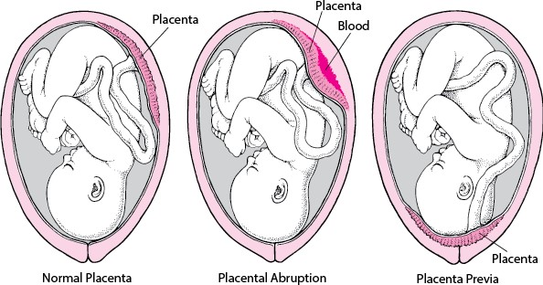
Choice B Reason: Placental abruption. This is a correct answer that explains the symptoms of fetal bradycardia, abdominal pain, and vaginal bleeding in a woman with a history of crack cocaine use. Placental abruption. This is because placental abruption is a condition where the placenta separates from the uterine wall before delivery, which can cause fetal distress, maternal hemorrhage, and shock. Placental abruption can be triggered by maternal hypertension, trauma, or substance abuse, such as crack cocaine.
Choice C Reason: Anaphylactoid syndrome of pregnancy. This is an incorrect answer that refers to a rare and fatal condition also known as amniotic fluid embolism. Anaphylactoid syndrome of pregnancy is a condition where amniotic fluid enters into the maternal bloodstream and causes an allergic reaction, which can lead to respiratory failure, cardiac arrest, coagulopathy, and coma. Anaphylactoid syndrome of pregnancy does not cause fetal bradycardia or vaginal bleeding.
Choice D Reason: Placenta previa. This is an incorrect answer that indicates another placental disorder. Placenta previa is a condition where the placenta covers or is near the cervix, which can cause painless bright red bleeding during pregnancy or labor. Placenta previa does not cause fetal bradycardia or abdominal pain.
Correct Answer is A
Explanation
Choice A Reason: "Our baby's newborn rash is from this syndrome." This is because this statement by a parent indicates that additional teaching is required, as it shows a misunderstanding or confusion about FAS and its manifestations. FAS is a condition that occurs when a woman consumes alcohol during pregnancy, which can affect the development and function of various organs and systems in the fetus and child. FAS can cause physical, behavioral, and cognitive problems such as facial abnormalities, growth retardation, learning difficulties, and atention deficits. FAS does not cause newborn rash, which is a common and benign condition that affects many newborns regardless of maternal alcohol intake. Newborn rash is also known as erythema toxicum neonatorum or baby acne, which is characterized by small red bumps or pustules on the face, chest, or back that usually disappear within a few weeks.
Choice B Reason: "His face looks like it does due to this problem." This is a correct answer that indicates adequate understanding of FAS and its features. Facial abnormalities are one of the characteristic signs of FAS, which include small eye openings, thin upper lip, flat nasal bridge, and smooth philtrum (the groove between the nose and upper lip).
Choice C Reason: "He can show signs of withdrawal from alcohol exposure like jiteriness, sweating, hyper reflexes, poor feeding and not sleeping well." This is a correct answer that indicates adequate understanding of FAS and its complications. Signs of withdrawal are possible effects of FAS, which occur when the fetus or newborn is exposed to alcohol in utero or through breast milk, which can cause neurotoxicity and dependency. Signs of withdrawal can include jiteriness, sweating, hyper reflexes, poor feeding, and not sleeping well, as well as irritability, seizures, or tremors.
Choice D Reason: "He is at risk of having intellectual disabilities, so we will need to get extra services to support him." This is a correct answer that indicates adequate understanding of FAS and its implications. Intellectual disabilities are potential outcomes of FAS, which affect the cognitive development and function of the child. Intellectual disabilities can cause problems with memory, Reasoning, language, and social skills. Extra services and support may be needed to help the child achieve their optimal potential and quality of life.
Whether you are a student looking to ace your exams or a practicing nurse seeking to enhance your expertise , our nursing education contents will empower you with the confidence and competence to make a difference in the lives of patients and become a respected leader in the healthcare field.
Visit Naxlex, invest in your future and unlock endless possibilities with our unparalleled nursing education contents today