Identify the structure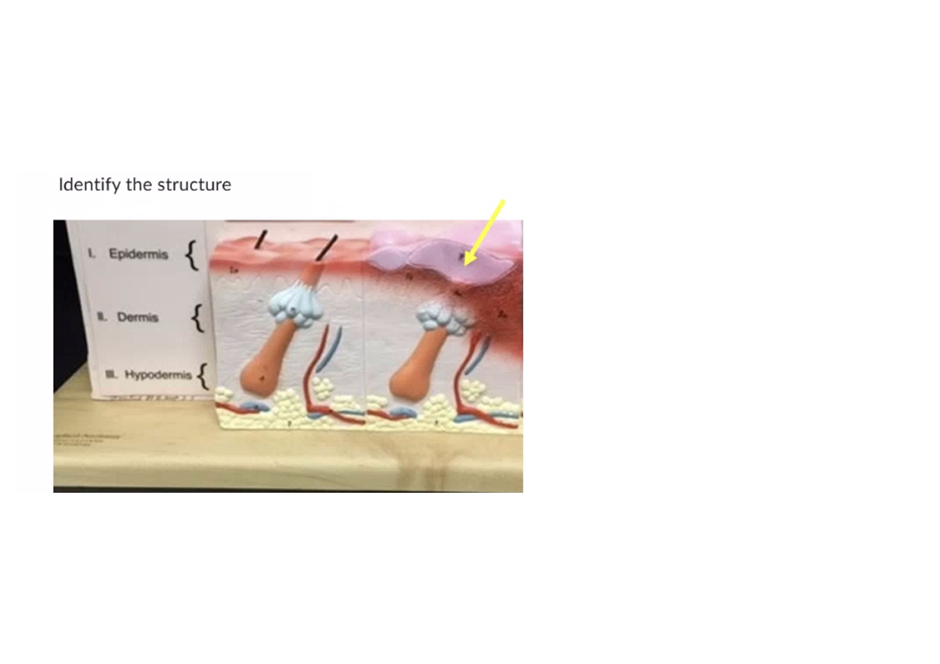
Stratum corneum
Stratum basale
Dermal papillae
Sebaceous gland
The Correct Answer is A
A. Stratum corneum: The stratum corneum is composed of dead, keratinized cells that form a tough, protective barrier. Its main function is to prevent water loss, protect against mechanical injury, and serve as the first line of defense against pathogens. Because it is the most superficial layer of the epidermis, it is the correct structure indicated by the arrow.
B. Stratum basale: This is the deepest layer of the epidermis, responsible for cell division and regeneration, not the outermost layer.
C. Dermal papillae: These are found in the dermis, not the epidermis. They interlock with the epidermis to strengthen the connection between the two layers.
D. Sebaceous gland: This gland is located in the dermis, associated with hair follicles, and secretes sebum. It is not part of the epidermis.
Nursing Test Bank
Naxlex Comprehensive Predictor Exams
Related Questions
Correct Answer is D
Explanation
A. Simple squamous epithelium: Simple squamous epithelium consists of a single layer of flat cells, specialized for diffusion and filtration (e.g., alveoli, capillaries). It does not provide the protective layering needed in the vaginal canal.
B. Stratified squamous epithelium (non-keratinized): The vaginal canal is lined with non-keratinized stratified squamous epithelium. This multilayered tissue provides protection against friction and mechanical stress during intercourse and childbirth, while remaining moist and flexible.
C. Transitional epithelium: Transitional epithelium is found in the urinary bladder and ureters, where it allows stretching. It is not present in the vaginal canal.
D. Adipose tissue: The tissue shown at the arrow in the anatomical model is adipose tissue (commonly known as fat).The yellow, pebbled, or "honeycomb" texture is the standard representation of fat deposits on medical models.
Correct Answer is A
Explanation
A. Glycolysis: This is the first stage of cellular respiration. As shown in the diagram, it occurs in the cytosol outside of the mitochondria and results in a small yield of ATP (indicated by the yellow starburst at the bottom).
B. Pyruvate Oxidation: This is the "transition" step represented by the smaller orange box where pyruvate enters the mitochondrial matrix to be converted into Acetyl-CoA.
C. Citric Acid Cycle (Krebs Cycle): This stage is represented by the red circular flow within the mitochondrial matrix, where electron carriers (NADH and $FADH_2$) are primarily generated.
D. Oxidative Phosphorylation: Indicated by the purple box on the right, this final stage occurs on the inner mitochondrial membrane (cristae) and produces the largest amount of ATP using the electron transport chain and chemiosmosis.
Whether you are a student looking to ace your exams or a practicing nurse seeking to enhance your expertise , our nursing education contents will empower you with the confidence and competence to make a difference in the lives of patients and become a respected leader in the healthcare field.
Visit Naxlex, invest in your future and unlock endless possibilities with our unparalleled nursing education contents today