Specifically, what type of tissue is shown here?
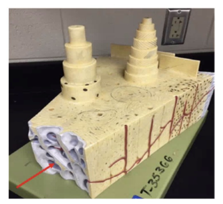
Compact Bone
Spongy (Cancellous) Bone
Hyaline Cartilage
Periosteum
The Correct Answer is B
A. Compact Bone: This is the dense, outer layer of bone organized into subunits called osteons (visible as the tall "towers" on top of the model). The arrow points to a different, porous region.
B. Spongy (Cancellous) Bone: The arrow points to the honeycomb-like network of bone called trabeculae. This tissue is found at the ends of long bones and lining the medullary cavity, providing structural support while keeping the skeleton lightweight.
C. Hyaline Cartilage: While often found at the ends of bones, hyaline cartilage is a smooth, glass-like connective tissue, not the porous, mineralized bone tissue shown here.
D. Periosteum: This is the fibrous membrane that covers the outer surface of bones. The arrow is pointing deep into the internal structure of the bone rather than the external surface.
Nursing Test Bank
Naxlex Comprehensive Predictor Exams
Related Questions
Correct Answer is B
Explanation
A. Cardiac Muscle Tissue: This tissue is found exclusively in the walls of the heart to facilitate involuntary rhythmic contractions. It is not found in the limbs.
B. Skeletal Muscle Tissue: This tissue is attached to bones and is responsible for voluntary movement. The arrow points to the quadriceps femoris group, which is composed of skeletal muscle fibers.
C. Fibrocartilage: While present in the knee (as the menisci), fibrocartilage is a tough, shock-absorbing tissue found between bones, not in the large, reddish contractile mass indicated by the arrow.
D. Dense Regular Connective Tissue: This tissue forms tendons and ligaments. While a tendon (the patellar tendon/ligament) is visible just below the arrow, the arrow specifically points to the belly of the muscle.
Correct Answer is D
Explanation
- G1 phase: This is the first gap phase where the cell grows and performs normal functions, not division.
- S phase: DNA replication occurs here, but chromosomes are not yet dividing.
- G2 phase: The cell prepares for mitosis by synthesizing proteins and checking DNA integrity, but division has not started.
- M phase (Mitosis): The cell undergoes nuclear division (prophase, metaphase, anaphase, telophase) followed by cytokinesis. The diagram highlights "A" in the central red segment, which typically represents mitosis in cell cycle charts. This is the stage where the cell actually divides, making it distinct from the preparatory phases.
Whether you are a student looking to ace your exams or a practicing nurse seeking to enhance your expertise , our nursing education contents will empower you with the confidence and competence to make a difference in the lives of patients and become a respected leader in the healthcare field.
Visit Naxlex, invest in your future and unlock endless possibilities with our unparalleled nursing education contents today