What stage of the cell cycle is shown here?
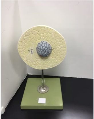
Prophase
Interphase
Metaphase
Telophase
The Correct Answer is B
- Prophase: Chromatin condenses into visible chromosomes, and the nuclear envelope begins to break down. This is not seen in the model.
- Interphase: During interphase, the cell is not actively dividing but is preparing for mitosis. The nuclear envelope remains intact, and the genetic material is visible as uncondensed chromatin rather than distinct chromosomes. This stage is crucial for DNA replication and cellular growth, ensuring the cell is ready for division. The presence of a clearly defined nucleus with diffuse chromatin confirms that the cell is in interphase
- Metaphase: Chromosomes align at the cell’s equatorial plate, which is absent here.
- Telophase: Chromosomes decondense, and two nuclear envelopes reform, indicating the end of mitosis. The model does not show this.
Nursing Test Bank
Naxlex Comprehensive Predictor Exams
Related Questions
Correct Answer is B
Explanation
A. DNA: DNA contains the bases adenine (A), thymine (T), cytosine (C), and guanine (G). The presence of uracil (U) in the model rules out DNA, since uracil is unique to RNA.
B. RNA: The model shows nucleotides labeled G (guanine), U (uracil), and C (cytosine), which are components of RNA. The entire structure represents a strand of RNA being synthesized or processed.
C. Protein: Proteins are composed of amino acids, not nucleotide bases. The colored blocks labeled with letters (G, U, C) clearly indicate nucleotides, not amino acids.
D. Transfer RNA (tRNA): While tRNA is a type of RNA, the model depicts a linear strand of RNA bases rather than the folded cloverleaf structure characteristic of tRNA.
Correct Answer is A
Explanation
A. Stratum corneum: The stratum corneum is composed of dead, keratinized cells that form a tough, protective barrier. Its main function is to prevent water loss, protect against mechanical injury, and serve as the first line of defense against pathogens. Because it is the most superficial layer of the epidermis, it is the correct structure indicated by the arrow.
B. Stratum basale: This is the deepest layer of the epidermis, responsible for cell division and regeneration, not the outermost layer.
C. Dermal papillae: These are found in the dermis, not the epidermis. They interlock with the epidermis to strengthen the connection between the two layers.
D. Sebaceous gland: This gland is located in the dermis, associated with hair follicles, and secretes sebum. It is not part of the epidermis.
Whether you are a student looking to ace your exams or a practicing nurse seeking to enhance your expertise , our nursing education contents will empower you with the confidence and competence to make a difference in the lives of patients and become a respected leader in the healthcare field.
Visit Naxlex, invest in your future and unlock endless possibilities with our unparalleled nursing education contents today