Where is the most ATP made?
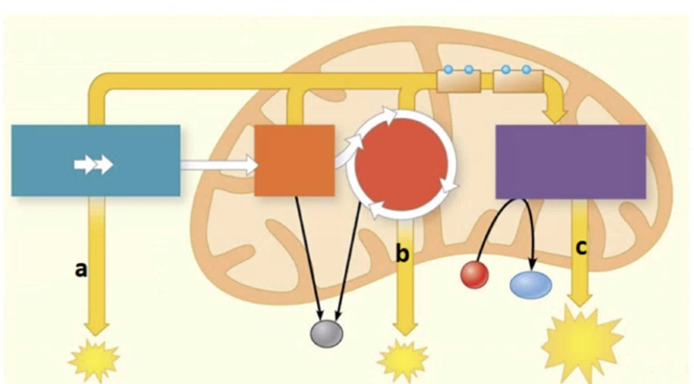
Stage a
Stage b
Stage c
ATP yield is equal in all stages
The Correct Answer is C
A. Stage a: This represents Glycolysis, which occurs in the cytosol and produces only a small net gain of 2 ATP per glucose molecule, represented by the small starburst.
B. Stage b: This represents the Citric Acid Cycle, which occurs in the mitochondrial matrix and also produces a relatively small amount of ATP (2 ATP) directly.
C. Stage c: This represents Oxidative Phosphorylation (Electron Transport Chain and Chemiosmosis). As indicated by the largest starburst in the diagram, this stage produces the vast majority of the cell's ATP (approximately 26-28 ATP).
D. ATP yield is equal: This is incorrect, as the chemical processes in the mitochondria are significantly more efficient at generating ATP than anaerobic glycolysis or the citric acid cycle alone.
Nursing Test Bank
Naxlex Comprehensive Predictor Exams
Related Questions
Correct Answer is B
Explanation
- Prophase: Chromatin condenses into visible chromosomes, and the nuclear envelope begins to break down. This is not seen in the model.
- Interphase: During interphase, the cell is not actively dividing but is preparing for mitosis. The nuclear envelope remains intact, and the genetic material is visible as uncondensed chromatin rather than distinct chromosomes. This stage is crucial for DNA replication and cellular growth, ensuring the cell is ready for division. The presence of a clearly defined nucleus with diffuse chromatin confirms that the cell is in interphase
- Metaphase: Chromosomes align at the cell’s equatorial plate, which is absent here.
- Telophase: Chromosomes decondense, and two nuclear envelopes reform, indicating the end of mitosis. The model does not show this.
Correct Answer is B
Explanation
A. Compact Bone: This is the dense, outer layer of bone organized into subunits called osteons (visible as the tall "towers" on top of the model). The arrow points to a different, porous region.
B. Spongy (Cancellous) Bone: The arrow points to the honeycomb-like network of bone called trabeculae. This tissue is found at the ends of long bones and lining the medullary cavity, providing structural support while keeping the skeleton lightweight.
C. Hyaline Cartilage: While often found at the ends of bones, hyaline cartilage is a smooth, glass-like connective tissue, not the porous, mineralized bone tissue shown here.
D. Periosteum: This is the fibrous membrane that covers the outer surface of bones. The arrow is pointing deep into the internal structure of the bone rather than the external surface.
Whether you are a student looking to ace your exams or a practicing nurse seeking to enhance your expertise , our nursing education contents will empower you with the confidence and competence to make a difference in the lives of patients and become a respected leader in the healthcare field.
Visit Naxlex, invest in your future and unlock endless possibilities with our unparalleled nursing education contents today