The arrow is pointing to what type of tissue?
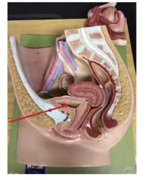
Simple Columnar Epithelium
Stratified Squamous Epithelium
Transitional Epithelium
Pseudostratified Ciliated Columnar Epithelium
The Correct Answer is C
A. Simple Columnar Epithelium: This tissue is typically found in areas involving high levels of absorption or secretion, such as the lining of the digestive tract (stomach and intestines). It does not have the elastic properties required for the urinary bladder.
B. Stratified Squamous Epithelium: This tissue consists of multiple layers of flat cells and is designed primarily for protection against friction and abrasion. It is found in the skin (epidermis), mouth, and esophagus. While durable, it lacks the specific distensibility characteristic of the bladder lining.
C. Transitional Epithelium: The urinary bladder requires a specialized lining that can withstand significant stretching as the organ fills with urine and recoil as it empties. Transitional epithelium (also known as urothelium) is uniquely designed for this; its cells change shape from rounded (cuboidal) when the bladder is empty to flattened (squamous-like) when the bladder is distended.
D. Pseudostratified Ciliated Columnar Epithelium: This tissue appears layered but is actually a single layer of cells of varying heights. It is almost exclusively found in the respiratory tract (trachea and bronchi), where cilia function to move mucus and trapped particles out of the airways.
Nursing Test Bank
Naxlex Comprehensive Predictor Exams
Related Questions
Correct Answer is B
Explanation
A. Hair Follicles: These are the tube-like structures that house the hair root, seen extending deep into the dermis in the model, but they are not the "humps" at the interface.
B. Dermal Papillae: These are the finger-like projections of the dermis that indent the overlying epidermis. They increase surface area for nutrient exchange and form the basis for fingerprints.
C. Sebaceous Glands: These are oil-producing glands typically associated with hair follicles, visible as multi-lobed structures in the dermis, but they are not the "hump" structures indicated.
D. Hypodermal Ridges: The hypodermis is the deep fatty layer (yellow tissue at the bottom). Ridges are not a standard anatomical term for structures at that specific epidermal-dermal junction.
Correct Answer is B
Explanation
A. Cardiac Muscle Tissue: This tissue is found exclusively in the walls of the heart to facilitate involuntary rhythmic contractions. It is not found in the limbs.
B. Skeletal Muscle Tissue: This tissue is attached to bones and is responsible for voluntary movement. The arrow points to the quadriceps femoris group, which is composed of skeletal muscle fibers.
C. Fibrocartilage: While present in the knee (as the menisci), fibrocartilage is a tough, shock-absorbing tissue found between bones, not in the large, reddish contractile mass indicated by the arrow.
D. Dense Regular Connective Tissue: This tissue forms tendons and ligaments. While a tendon (the patellar tendon/ligament) is visible just below the arrow, the arrow specifically points to the belly of the muscle.
Whether you are a student looking to ace your exams or a practicing nurse seeking to enhance your expertise , our nursing education contents will empower you with the confidence and competence to make a difference in the lives of patients and become a respected leader in the healthcare field.
Visit Naxlex, invest in your future and unlock endless possibilities with our unparalleled nursing education contents today