Name the structure labeled #3 on the following image:
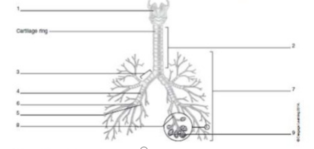
Primary bronchus
Respiratory bronchiole
Trachea
Secondary bronchus
The Correct Answer is A
A. Primary bronchus: The primary (main) bronchus is the first large airway branch that arises at the carina from the trachea and conducts air into each lung.
B. Respiratory bronchiole: Respiratory bronchioles are very small distal airways that contain alveoli and are deep within the lung parenchyma; they are much smaller than the structure shown near the main branch.
C. Trachea: The trachea is the central airway above the bifurcation (a vertical tube with cartilage rings); a label on the long central tube would indicate the trachea, not the branching airway.
D. Secondary bronchus: Secondary (lobar) bronchi are branches of the primary bronchus that lead to lung lobes; they are a generation distal to the main bronchus and are smaller than the primary bronchus.
Nursing Test Bank
Naxlex Comprehensive Predictor Exams
Related Questions
Correct Answer is D
Explanation
A. Glomerulus:The glomerulus is the filtration site; ADH/aldosterone act downstream on tubular epithelial cells, not on glomerular filtration directly.
B. Loop of Henle: The loop establishes the medullary gradient; ADH/aldosterone have limited direct action here (ADH affects water permeability mainly in collecting ducts; aldosterone acts mainly on DCT/collecting duct).
C. Vasa recta:Vasa recta are blood vessels that help preserve the medullary gradient (countercurrent exchange) but are not the primary hormone-target epithelial segments for ADH/aldosterone.
D. DCT and collecting duct:Aldosterone acts on the distal convoluted tubule and cortical collecting duct to increase Na⁺ reabsorption (and K⁺ secretion); ADH acts on the late DCT and collecting duct to increase water reabsorption by inserting aquaporins -together they adjust water and sodium retention.
Correct Answer is C
Explanation
A. Uterus: The uterus is an internal reproductive organ (the womb) and does not include the labia.
B. Fallopian tube: Fallopian (uterine) tubes are internal conduits for the oocyte; they are not the labia.
C. Vulva:The vulva is the external female genitalia and includes the labia majora and labia minora .
D. Ovary: The ovaries are internal gonads that produce oocytes and hormones; they are not the labia.
Whether you are a student looking to ace your exams or a practicing nurse seeking to enhance your expertise , our nursing education contents will empower you with the confidence and competence to make a difference in the lives of patients and become a respected leader in the healthcare field.
Visit Naxlex, invest in your future and unlock endless possibilities with our unparalleled nursing education contents today