The nurse examines a client's auditory canal and tympanic membrane with an otoscope. The nurse recognizes that which of the following is considered an abnormal finding?
A shiny, pearly white color tympanic membrane
The presence of cerumen
The presence of a cone of light
A yellow or amber color to the tympanic membrane.
The Correct Answer is D
A. A shiny, pearly white color tympanic membrane: This is a normal finding. A healthy tympanic membrane often appears shiny and pearly white.
B. The presence of cerumen: This is a normal finding. Cerumen, or earwax, is a natural substance that helps protect the ear canal.
C. The presence of a cone of light: This is a normal finding. The cone of light is a reflection of the otoscope light on the tympanic membrane and is a normal variation.
D. A yellow or amber color to the tympanic membrane: This is considered an abnormal finding. A yellow or amber coloration of the tympanic membrane can indicate the presence of fluid or infection behind the eardrum, which may be a sign of otitis media or other ear conditions.

Nursing Test Bank
Naxlex Comprehensive Predictor Exams
Related Questions
Correct Answer is C
Explanation
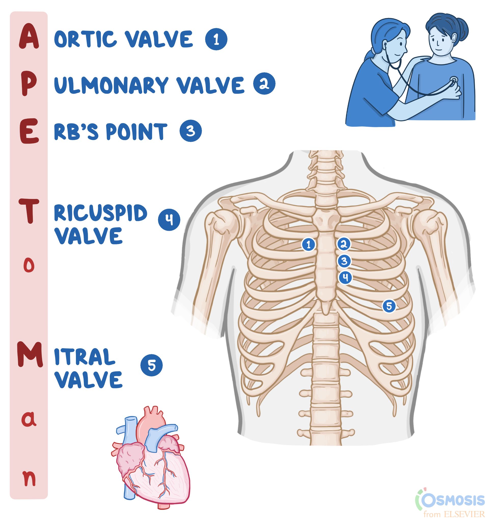
A. Fifth intercostal space, left of the midclavicular line: This placement is used to auscultate the mitral valve, which is best heard at the apex of the heart. The mitral valve sounds are typically heard around the fifth intercostal space, midclavicular line.
B. Left lower sternal border: This placement is used to auscultate the tricuspid valve, which is best heard at the lower left sternal border.
C. Second left intercostal space: This is the correct placement for auscultating the pulmonic valve. The pulmonic valve sounds are best heard at the second left intercostal space, which is close to the upper left sternal border.
D. Second right intercostal space: This placement is used to auscultate the aortic valve, which is best heard at the second right intercostal space, close to the upper right sternal border.
Correct Answer is A
Explanation
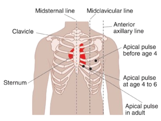
A. Fifth left intercostal space at the midclavicular line:
Explanation: The apical pulse, or the point of maximal impulse (PMI), is typically located at the fifth intercostal space at the midclavicular line on the chest. This is the area where the heartbeat is best heard using a stethoscope in most adults.
B. Third left intercostal space at the midclavicular line:
Explanation: This location is too high for the apical pulse. The heart's apex is generally not found at the third intercostal space; it's lower, closer to the fifth intercostal space.
C. Fourth left intercostal space at the sternal border:
Explanation: This location is not the typical site for auscultating the apical pulse. The PMI is usually heard at the midclavicular line, not at the sternal border.
D. Under the left breast at the midclavicular line:
Explanation: This position is not precise enough for auscultating the apical pulse. The specific intercostal space (fifth) and midclavicular line are crucial for accurate assessment.
Whether you are a student looking to ace your exams or a practicing nurse seeking to enhance your expertise , our nursing education contents will empower you with the confidence and competence to make a difference in the lives of patients and become a respected leader in the healthcare field.
Visit Naxlex, invest in your future and unlock endless possibilities with our unparalleled nursing education contents today