The nurse is discussing the blood flow pattern of the heart. The nurse should recognize which of the following as the accurate blood flow pattern of the heart?
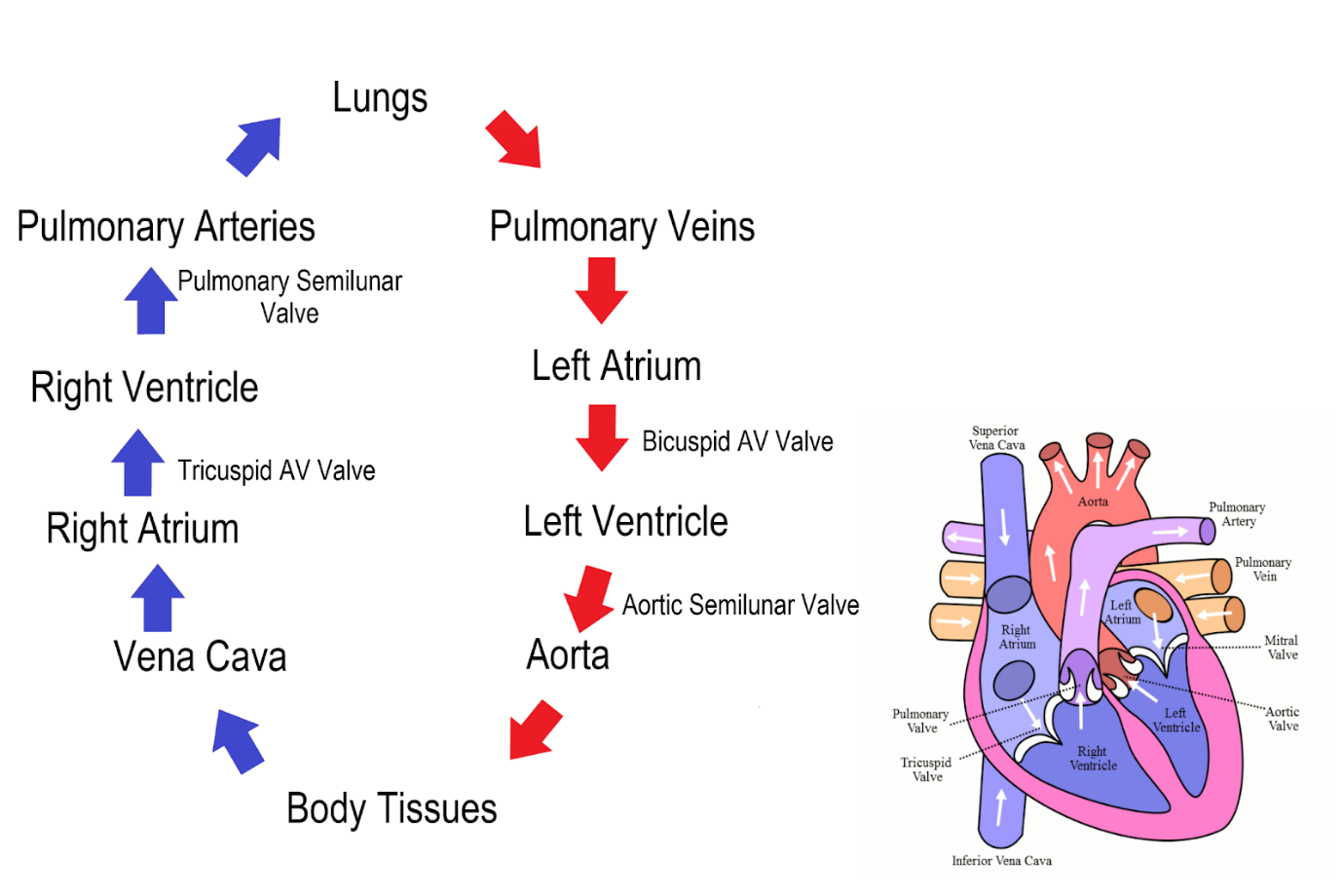
Aorta-Right atrium-right ventricle-pulmonary vein-lungs- pulmonary artery left atrium-left ventricle
vena cava-right atrium-right ventricle-pulmonary vein-lungs- pulmonary artery-left atrium-left ventricle
vena cava→ right atrium-right ventricle-pulmonary artery-lungs- pulmonary vein-left atrium-left ventricle
Aorta-right atrium-right ventricle-lungs-pulmonary vein-left atrium-left ventricle- vena cava
The Correct Answer is C
In this sequence:
Vena cava brings deoxygenated blood from the body into the right atrium.
Blood flows from the right atrium to the right ventricle through the tricuspid valve.
The right ventricle pumps blood into the pulmonary artery to the lungs.
In the lungs, blood is oxygenated and returns to the heart via the pulmonary veins into the left atrium.
From the left atrium, blood moves to the left ventricle through the bicuspid (mitral) valve.
The left ventricle then pumps oxygenated blood into the body through the aorta.
This sequence represents the systemic and pulmonary circulation of the heart.
Nursing Test Bank
Naxlex Comprehensive Predictor Exams
Related Questions
Correct Answer is C
Explanation
A. Checks the instrument gauge to ensure the reading starts at zero:
This action is correct. Before taking a blood pressure reading, it's essential to ensure that the instrument's gauge starts at zero. This ensures accurate measurement as the reading reflects the pressure above zero.
B. Centers the cuff bladder over the client's brachial artery:
This action is correct. Proper placement of the blood pressure cuff over the brachial artery is crucial for accurate readings. Centering the cuff ensures that the artery is correctly compressed for measurement.
C. Places the client's arm above the level of the client's heart:
This action is incorrect. Placing the arm above heart level can result in a falsely low blood pressure reading. The arm should be at the same level as the heart to obtain an accurate measurement.
D. Wraps the blood pressure cuff around the client's arm using firm pressure:
This action is correct, but it's important to note that while the cuff should be snug, it should not be too tight or too loose. Wrapping the cuff with firm, even pressure ensures proper compression of the artery for an accurate measurement.
Correct Answer is A
Explanation
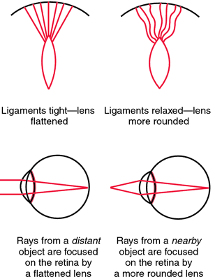
A. The pupils constrict when the examiner's index finger slowly moves toward the client's nose.
This statement is correct. Visual accommodation is the process by which the eye's lens changes shape to focus on objects at varying distances. When an object moves closer to the eyes, the pupils constrict to adjust and focus on the near object, preventing double vision.
B. The client involuntarily blinks in the presence of bright light directed over the pupils during the eye exam.
This statement describes the pupillary light reflex, not visual accommodation. The pupillary light reflex is the response of the pupils to light exposure.
C. The client's peripheral vision becomes sharper when the examiner shines a light over the pupils.
This statement is not accurate. Peripheral vision sharpness is not related to visual accommodation. Visual accommodation mainly involves adjusting focus for objects at varying distances.
D. The pupils dilate when the examiner's index finger slowly moves toward the client's nose.
This statement is incorrect. Pupils should constrict, not dilate, when focusing on a near object (as in visual accommodation). Dilation occurs in low-light conditions or in response to sympathetic stimulation.
Whether you are a student looking to ace your exams or a practicing nurse seeking to enhance your expertise , our nursing education contents will empower you with the confidence and competence to make a difference in the lives of patients and become a respected leader in the healthcare field.
Visit Naxlex, invest in your future and unlock endless possibilities with our unparalleled nursing education contents today