
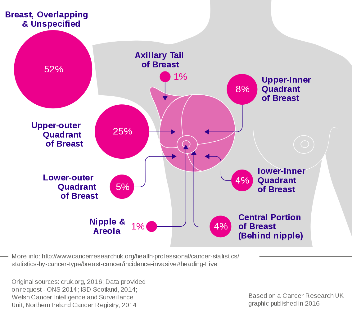
The nurse is performing a breast examination on a client. The nurse understands that examining the upper outer quadrant of the breast is especially important because this part is:
The location of most breast tumors.
More prone to Injury and calcifications than other locations in the breast
The largest quadrant of the breast
Where most of the suspensory ligaments attach
The Correct Answer is A
A. The location of most breast tumors: This choice is correct. The upper outer quadrant of the breast is the most common site for breast tumors. This area has a higher concentration of glandular tissue, making it more susceptible to the development of tumors. Therefore, thorough examination of this area is crucial during breast examinations.
B. More prone to Injury and calcifications than other locations in the breast: While injuries can occur in any part of the breast, the statement about calcifications is not accurate. Calcifications in breast tissue can occur in various areas and are not specific to the upper outer quadrant.
C. The largest quadrant of the breast: This statement is not accurate. The breast quadrants are not distinguished by size; they are anatomical divisions used for reference during breast examinations. Each quadrant has its significance for examination purposes, but none is considered the largest.
D. Where most of the suspensory ligaments attach: The suspensory ligaments, also known as Cooper's ligaments, provide structural support to the breast tissue. While they are essential for breast anatomy, they are not concentrated in the upper outer quadrant exclusively. These ligaments are distributed throughout the breast tissue.
Nursing Test Bank
Naxlex Comprehensive Predictor Exams
Related Questions
Correct Answer is D
Explanation
A. Percussion of the posterior chest: Percussion helps assess the underlying structures of the chest but does not directly confirm symmetric chest expansion.
B. Inspection of the shape and configuration of the chest wall: Inspection is a crucial part of assessing chest symmetry. Any deformities, asymmetry, or abnormalities in the shape and configuration of the chest wall can be visually identified.
C. Placing the palmar surface of the fingers of one hand against the chest and having the client repeat "ninety-nine": This technique, known as tactile fremitus, involves feeling for vibrations or tremors while the client repeats a phrase. While it can provide information about underlying lung conditions, it's not primarily used to confirm symmetric chest expansion.
D. Placing hands sideways on the posterolateral chest wall with thumbs pointing together at the level of T9 or T10: This technique, known as chest expansion measurement, is used to assess symmetric chest expansion. Placing hands in this manner allows the nurse to feel for bilateral chest expansion during inspiration, ensuring that both sides of the chest expand symmetrically.

Correct Answer is C
Explanation
A. Bronchovesicular breath sounds and normal in that location:
Bronchovesicular breath sounds are medium-pitched sounds heard over the major bronchi and are usually equal on inspiration and expiration. They are typically heard in the 1st and 2nd intercostal spaces anteriorly and between the scapulae posteriorly. While they might be normal in certain locations, hearing them over peripheral lung fields might indicate an abnormality.
B. Normally auscultated over the trachea:
This statement doesn't specify a particular type of breath sound. Tracheal breath sounds are harsh and relatively high-pitched, heard directly over the trachea. They are normal over the trachea but are not normally heard in the lung periphery.
C. Vesicular breath sounds and normal in that location:
Vesicular breath sounds are low-pitched, soft sounds heard over most of the lungs during inspiration. They are longer on inspiration than expiration and are considered normal breath sounds heard in the peripheral lung fields. Hearing vesicular sounds in the posterior lower lobes is typical and indicates normal lung function.
D. Bronchial breath sounds and normal in that location:
Bronchial breath sounds are high-pitched and loud, heard primarily over the trachea and larynx. If heard in the peripheral lung fields, especially in the lower lobes, it can suggest an abnormality such as consolidation or compression of lung tissue.
Whether you are a student looking to ace your exams or a practicing nurse seeking to enhance your expertise , our nursing education contents will empower you with the confidence and competence to make a difference in the lives of patients and become a respected leader in the healthcare field.
Visit Naxlex, invest in your future and unlock endless possibilities with our unparalleled nursing education contents today