What structures are shown here? "The Humps"
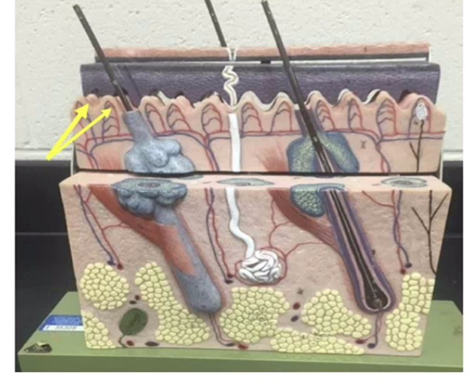
Hair Follicles
Dermal Papillae
Sebaceous Glands
Hypodermal Ridges
The Correct Answer is B
A. Hair Follicles: These are the tube-like structures that house the hair root, seen extending deep into the dermis in the model, but they are not the "humps" at the interface.
B. Dermal Papillae: These are the finger-like projections of the dermis that indent the overlying epidermis. They increase surface area for nutrient exchange and form the basis for fingerprints.
C. Sebaceous Glands: These are oil-producing glands typically associated with hair follicles, visible as multi-lobed structures in the dermis, but they are not the "hump" structures indicated.
D. Hypodermal Ridges: The hypodermis is the deep fatty layer (yellow tissue at the bottom). Ridges are not a standard anatomical term for structures at that specific epidermal-dermal junction.
Nursing Test Bank
Naxlex Comprehensive Predictor Exams
Related Questions
Correct Answer is B
Explanation
A. DNA: DNA contains the bases adenine (A), thymine (T), cytosine (C), and guanine (G). The presence of uracil (U) in the model rules out DNA, since uracil is unique to RNA.
B. RNA: The model shows nucleotides labeled G (guanine), U (uracil), and C (cytosine), which are components of RNA. The entire structure represents a strand of RNA being synthesized or processed.
C. Protein: Proteins are composed of amino acids, not nucleotide bases. The colored blocks labeled with letters (G, U, C) clearly indicate nucleotides, not amino acids.
D. Transfer RNA (tRNA): While tRNA is a type of RNA, the model depicts a linear strand of RNA bases rather than the folded cloverleaf structure characteristic of tRNA.
Correct Answer is B
Explanation
A. Compact Bone: This is the dense, outer layer of bone organized into subunits called osteons (visible as the tall "towers" on top of the model). The arrow points to a different, porous region.
B. Spongy (Cancellous) Bone: The arrow points to the honeycomb-like network of bone called trabeculae. This tissue is found at the ends of long bones and lining the medullary cavity, providing structural support while keeping the skeleton lightweight.
C. Hyaline Cartilage: While often found at the ends of bones, hyaline cartilage is a smooth, glass-like connective tissue, not the porous, mineralized bone tissue shown here.
D. Periosteum: This is the fibrous membrane that covers the outer surface of bones. The arrow is pointing deep into the internal structure of the bone rather than the external surface.
Whether you are a student looking to ace your exams or a practicing nurse seeking to enhance your expertise , our nursing education contents will empower you with the confidence and competence to make a difference in the lives of patients and become a respected leader in the healthcare field.
Visit Naxlex, invest in your future and unlock endless possibilities with our unparalleled nursing education contents today