What type of burn is shown here?
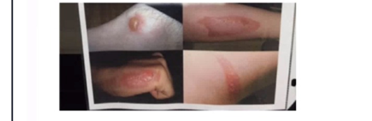
First-degree burn
Second-degree burn
Third-degree burn
Fourth-degree burn
The Correct Answer is B
A. First-degree burn: First-degree burns involve only the epidermis, presenting with redness and pain but no blistering. The images show blister formation and deeper tissue involvement, which rules out first-degree.
B. Second-degree burn: Second-degree burns extend into the dermis and are characterized by blistering, redness, swelling, and significant pain. The burns in the images display these hallmark features, making this the best answer.
C. Third-degree burn: Third-degree burns destroy the entire epidermis and dermis, often appearing white, charred, or leathery, and may be painless due to nerve damage. The images do not show this level of tissue destruction.
D. Fourth-degree burn: Fourth-degree burns extend beyond the skin into muscle, bone, or deeper structures. These are extremely severe and not represented in the images provided.
Nursing Test Bank
Naxlex Comprehensive Predictor Exams
Related Questions
Correct Answer is C
Explanation
A. Stage a: This represents Glycolysis, which occurs in the cytosol and produces only a small net gain of 2 ATP per glucose molecule, represented by the small starburst.
B. Stage b: This represents the Citric Acid Cycle, which occurs in the mitochondrial matrix and also produces a relatively small amount of ATP (2 ATP) directly.
C. Stage c: This represents Oxidative Phosphorylation (Electron Transport Chain and Chemiosmosis). As indicated by the largest starburst in the diagram, this stage produces the vast majority of the cell's ATP (approximately 26-28 ATP).
D. ATP yield is equal: This is incorrect, as the chemical processes in the mitochondria are significantly more efficient at generating ATP than anaerobic glycolysis or the citric acid cycle alone.
Correct Answer is A
Explanation
A. Chromatid: In this chromosome model, the pink structure represents the chromatid, and the red coiled section highlights a specific portion of the DNA molecule.
B. Centromere: This is the constricted region of the chromosome that attaches to spindle fibers during cell division, not the highlighted locus.
C. Telomere: These are repetitive DNA sequences at the ends of chromosomes that protect against degradation, not the middle region.
D. Histone protein: These are proteins around which DNA winds to form nucleosomes, but they are not visible as a distinct locus on a chromosome model.
Whether you are a student looking to ace your exams or a practicing nurse seeking to enhance your expertise , our nursing education contents will empower you with the confidence and competence to make a difference in the lives of patients and become a respected leader in the healthcare field.
Visit Naxlex, invest in your future and unlock endless possibilities with our unparalleled nursing education contents today