Which of the following is the structure through which blood exits the glomerulus?
Efferent arteriole
Proximal tubule
Distal tubule
Afferent arteriole.
The Correct Answer is A
The glomerulus is the main filtering unit of the kidney.
It is formed by a network of small blood vessels (capillaries) enclosed within a sac called the Bowman’s capsule.
The blood supply to the glomerulus is provided via the afferent arteriole.
The blood then flows through the capillary network, where it gets filtered, and then leaves the glomerulus via the efferent arteriole.
Choice B. Proximal tubule is not correct because it is where the ultrafiltrate collected in the Bowman’s space drains directly into.
Choice C. Distal tubule is not correct because it is not mentioned in relation to blood exiting the glomerulus.
Choice D. Afferent arteriole is not correct because it provides blood supply to the glomerulus.
Nursing Test Bank
Naxlex Comprehensive Predictor Exams
Related Questions
Correct Answer is C
Explanation
In this cross, both parents are homozygous recessive for the smooth leaf trait (ff).
This means that all of their offspring will inherit two copies of the recessive allele (f) and will therefore have smooth leaves.
Choice A. FF x FF is not correct because both parents are homozygous dominant for the fuzzy leaf trait (FF) and all of their offspring will inherit two copies of the dominant allele (F) and will therefore have fuzzy leaves.
Choice B. Ff x Ff is not correct because both parents are heterozygous for the leaf trait (Ff) and their offspring can inherit either one dominant allele (F) or one recessive allele (f) from each parent, resulting in a 3:1 ratio of fuzzy to smooth leaves.
Choice D. Ff x ff is not correct because one parent is heterozygous for the leaf trait (Ff) while the other is homozygous recessive (ff), resulting in a 1:1 ratio of fuzzy to smooth leaves in their offspring.
Correct Answer is B
Explanation
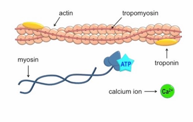
Calcium ions play a crucial role in initiating muscle contraction.
When a muscle cell is stimulated to contract by an action potential, calcium channels open in the sarcoplasmic membrane and release calcium into the sarcoplasm.
Some of this calcium attaches to troponin, which causes it to change shape. This shape change exposes binding sites for myosin on the actin filaments.
Myosin’s binding to actin causes cross-bridge formation, and muscle contraction begins.
The other ions mentioned in the question do not have this specific role in muscle contraction.
Potassium ions are important for maintaining the resting membrane potential of cells, but they do not bind to the troponin complex.
Phosphorus ions are important for energy metabolism but do not bind to the troponin complex.
Sodium ions are important for generating action potentials but do not bind to the troponin complex.
Whether you are a student looking to ace your exams or a practicing nurse seeking to enhance your expertise , our nursing education contents will empower you with the confidence and competence to make a difference in the lives of patients and become a respected leader in the healthcare field.
Visit Naxlex, invest in your future and unlock endless possibilities with our unparalleled nursing education contents today