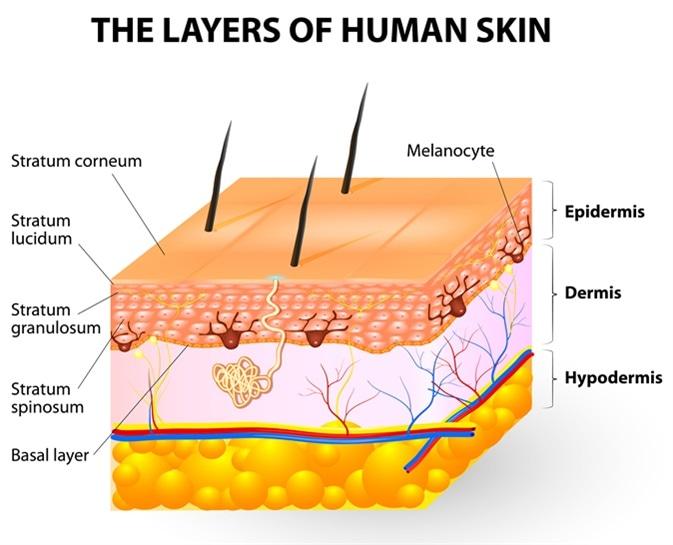
Melanocytes give rise to the pigment melanin, which is responsible for skin color. Where can the melanocytes be found?
Loose connective tissue
Epidermis
Dermis
Superficial fascia
The Correct Answer is B
A. Loose connective tissue:
Melanocytes are not typically found in loose connective tissue. Their primary location is within the epidermis, specifically in the basal layer, where they interact with keratinocytes to produce melanin and contribute to skin color. Loose connective tissue contains collagen and elastin fibers, as well as fibroblasts, but it does not house melanocytes.
B. Epidermis:
This is the correct answer. Melanocytes are primarily located in the basal layer of the epidermis, which is the deepest layer of the epidermis. These cells produce melanin, a pigment that helps protect the skin from UV radiation and determines skin color. Melanocytes are interspersed among keratinocytes in the epidermis and transfer melanin to keratinocytes to provide skin pigmentation.
C. Dermis:
The dermis is the layer of skin beneath the epidermis and consists of connective tissue, blood vessels, nerves, hair follicles, and sweat glands. While the dermis plays a crucial role in supporting and nourishing the epidermis, melanocytes are not primarily located in the dermis. They are confined to the basal layer of the epidermis.
D. Superficial fascia:
The superficial fascia, also known as the subcutaneous tissue or hypodermis, lies beneath the dermis and consists of adipose (fat) tissue and connective tissue. It provides insulation, energy storage, and cushioning for underlying structures. However, melanocytes are not typically found in the superficial fascia. They are restricted to the epidermis, specifically the basal layer, where they carry out their function of melanin production.
Nursing Test Bank
Naxlex Comprehensive Predictor Exams
Related Questions
Correct Answer is A
Explanation
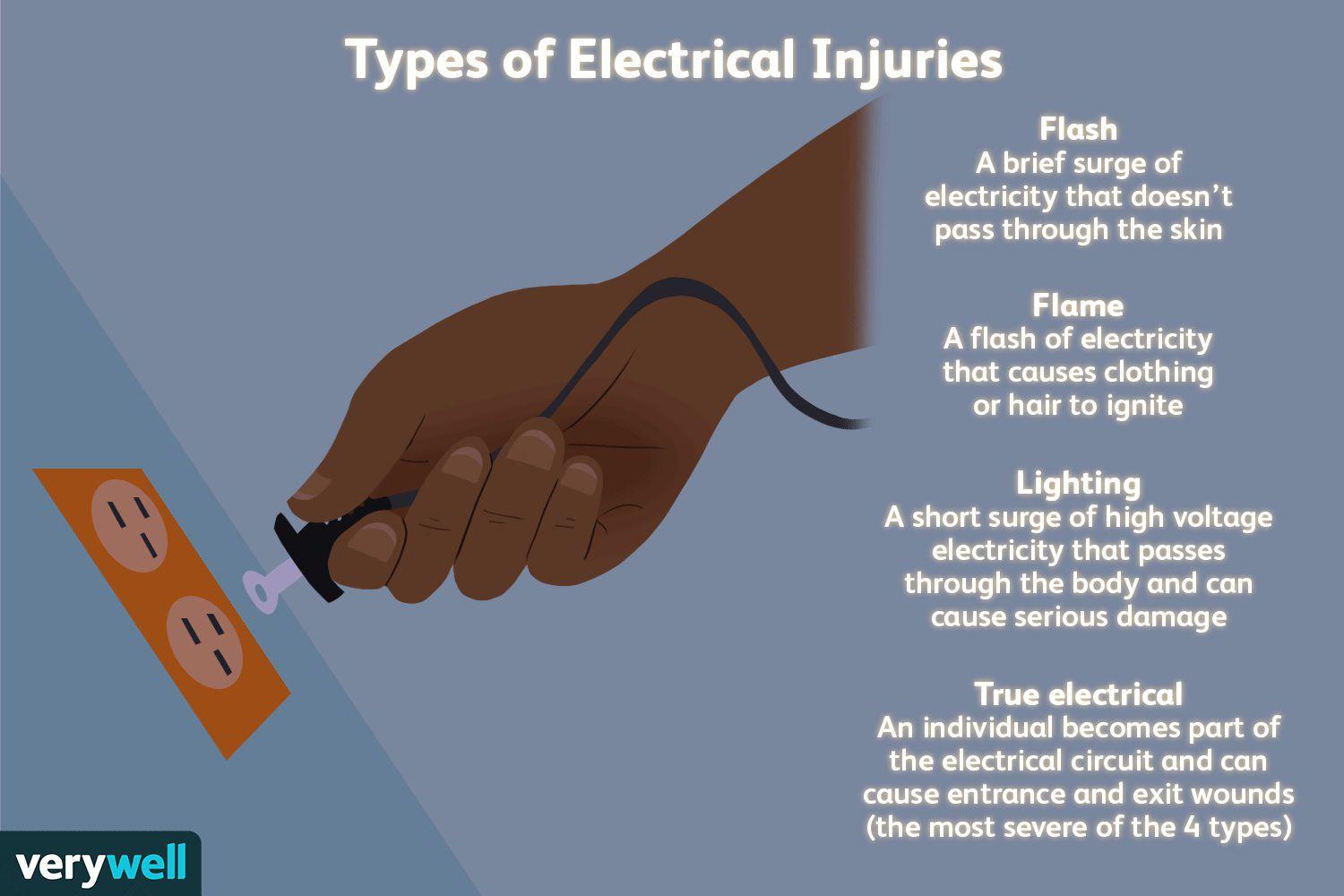
A. Electrical burns can have small amounts of skin damage, but more extensive damage beneath the skin.
This response is the best choice because it educates the client about the potential for deeper tissue damage associated with electrical burns. It acknowledges that while the burn on the skin may appear small, the damage underneath could be more extensive, affecting muscles, nerves, and blood vessels.
B. Electrical burns commonly cause reddened/purplish skin without blistering.
This statement is not the best response because it focuses solely on the appearance of the skin without addressing the potential for deeper tissue damage. While it is true that electrical burns can present with reddened or purplish skin without blistering, this response does not provide comprehensive information about the nature and severity of electrical burns.
C. Electrical burns typically are minor.
This response is incorrect because it downplays the seriousness of electrical burns. While some electrical burns may indeed be minor, others can cause significant tissue damage and complications. It's important for the nurse to educate the client about the range of severity that electrical burns can present.
D. Electrical burns usually cause much more skin damage than what can be seen on your skin.
This statement is partially accurate but does not provide as much information as choice A. While it acknowledges that electrical burns can cause more damage than what is visible on the skin's surface, it doesn't emphasize the potential for deeper tissue damage as effectively as choice A does.
Correct Answer is A
Explanation
A. The transfusion will begin after the administration of 650 mg of acetaminophen (Tylenol).
This option suggests that the nurse would administer acetaminophen to lower the client's temperature and then proceed with the blood transfusion. While acetaminophen can be used to reduce fever, the decision to administer medication should be made by the healthcare provider after assessing the client's overall condition and determining the cause of the fever. Administering medication without proper evaluation and orders from the healthcare provider is not appropriate.
B. The blood will be held, and the health care provider will be notified.
This option is the correct choice. When a client has an elevated temperature before a blood transfusion, it is standard practice to hold the transfusion and notify the healthcare provider. An elevated temperature could indicate an underlying infection or another condition that needs to be evaluated before proceeding with the transfusion to ensure the client's safety.
C. The transfusion will begin after the administration of an antihistamine.
Administering an antihistamine would not be the appropriate action in response to an elevated temperature before a blood transfusion. Antihistamines are typically used to treat allergic reactions, not fevers. Holding the transfusion and notifying the healthcare provider to assess the situation would be the correct course of action.
D. The transfusion will begin as prescribed.
This option is not appropriate because starting the transfusion without addressing the elevated temperature could pose risks to the client's health. Elevated temperatures may indicate an underlying infection or other conditions that need to be evaluated before proceeding with the transfusion. Holding the transfusion and seeking further guidance from the healthcare provider is the recommended action in this scenario.
Whether you are a student looking to ace your exams or a practicing nurse seeking to enhance your expertise , our nursing education contents will empower you with the confidence and competence to make a difference in the lives of patients and become a respected leader in the healthcare field.
Visit Naxlex, invest in your future and unlock endless possibilities with our unparalleled nursing education contents today