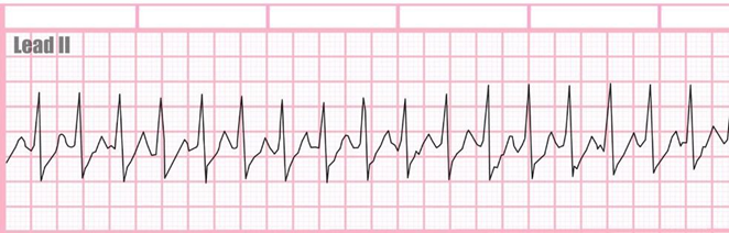
Mr Jones came to the ER complaining of chest palpitations. He states his heart feels like it is "racing". The nurse found the following rhythm on the monitor. What is the next step would the nurse expect to take first?
Defibrillate
Ask him to blow into a syringe
Draw troponins
Give amiodarone
The Correct Answer is B
B This maneuver, called the Valsalva maneuver, can sometimes help to restore normal heart rhythm in cases of SVT. It involves blowing forcefully into a syringe or performing a similar action that increases intra-abdominal pressure, which can stimulate the vagus nerve and help to slow down the heart rate.
A Defibrillation is a treatment used for life-threatening cardiac arrhythmias, particularly ventricular fibrillation or pulseless ventricular tachycardia. It involves delivering a therapeutic dose of electrical energy to the heart with a device called a defibrillator.
C Drawing troponin levels may be appropriate if there is suspicion of myocardial infarction as the cause of chest palpitations or if there are other symptoms suggestive of acute coronary syndrome. However, in the context of SVT presenting with chest palpitations and a racing heart, the priority is to address the arrhythmia first
D Amiodarone is an antiarrhythmic medication used to treat various types of cardiac arrhythmias, including ventricular and supraventricular arrhythmias. While it can be effective in certain cases of SVT, it is not typically the first-line treatment or the immediate next step in managing SVT in the emergency department.
Nursing Test Bank
Naxlex Comprehensive Predictor Exams
Related Questions
Correct Answer is B
Explanation
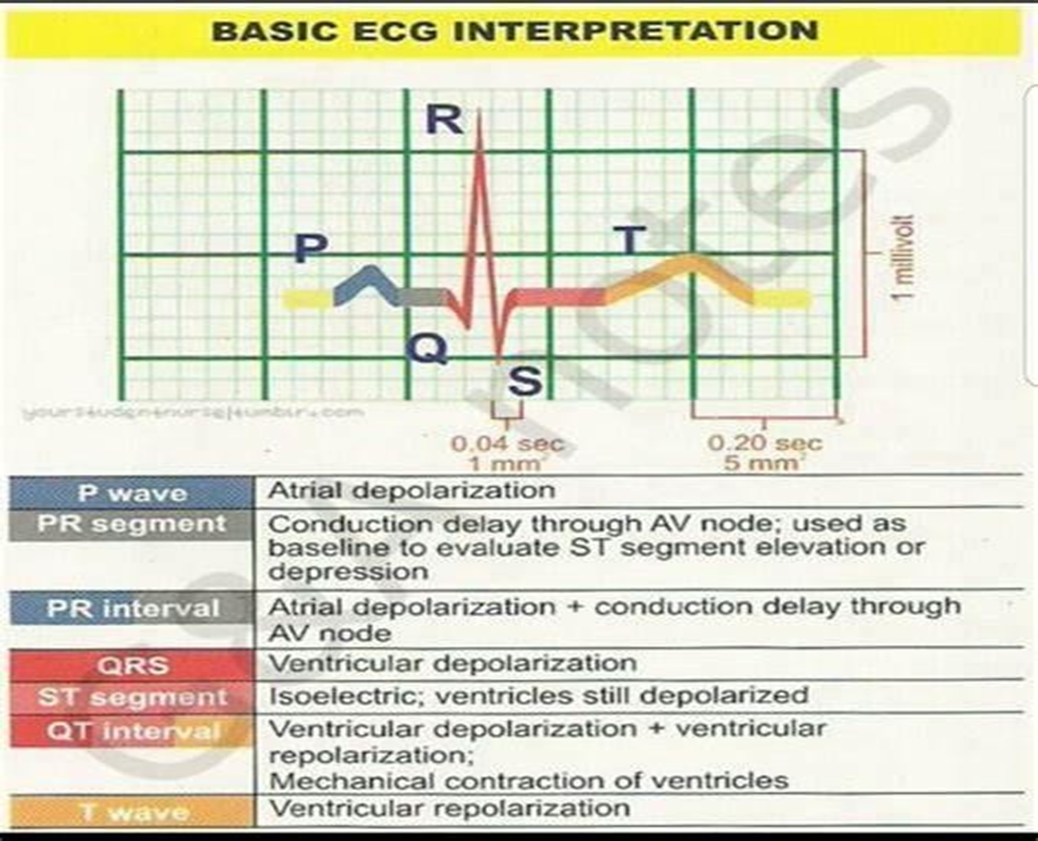
B. The QT interval represents ventricular depolarization and repolarization. It starts at the beginning of the QRS complex and ends at the end of the T wave. The QT interval reflects the total time it takes for both ventricular depolarization and repolarization to occur. Prolongation of the QT interval can be associated with an increased risk of arrhythmias, including torsades de pointes.
A. The QRS complex represents ventricular depolarization, which is the electrical activation of the ventricles. The duration of the QRS complex provides information about the time it takes for ventricular depolarization to occur. A prolonged QRS complex can indicate abnormalities in ventricular conduction, such as bundle branch blocks or ventricular hypertrophy.
C. The ST segment represents the early part of ventricular repolarization. It starts at the end of the QRS complex and ends at the beginning of the T wave. Changes in the ST segment, such as elevation or depression, can indicate myocardial ischemia or injury.
D. The PR interval represents the time it takes for the electrical impulse to travel from the atria to the ventricles. It includes atrial depolarization, atrial contraction, and the delay at the atrioventricular node. The PR interval does not specifically provide information about ventricular depolarization and repolarization.
Correct Answer is C
Explanation
C. Hypovolemia, or low blood volume, can lead to decreased venous return to the heart and reduced filling pressures. Consequently, CVP may decrease in hypovolemic states. Low CVP may indicate inadequate preload and reduced cardiac output, which are characteristic of hypovolemia.
A. Left ventricular failure typically results in elevated filling pressures rather than low CVP. In left ventricular failure, blood backs up into the pulmonary circulation, leading to increased pulmonary venous pressure and potentially elevated pulmonary capillary wedge pressure (PCWP), which is a surrogate marker for left atrial pressure. This elevated pressure is reflected in the CVP as well, resulting in increased CVP rather than low CVP.
B. Fluid overload typically results in elevated filling pressures and increased CVP rather than low CVP. Excess fluid volume increases venous return to the heart, leading to increased pressure within the central veins and elevated CVP.
D. Intracardiac shunts may cause alterations in cardiac pressures, but they typically do not result in consistently low CVP. Depending on the type and severity of the shunt, the direction and magnitude of pressure changes may vary. However, in the absence of other pathophysiological factors, intracardiac shunts are less likely to cause consistently low CVP.
Whether you are a student looking to ace your exams or a practicing nurse seeking to enhance your expertise , our nursing education contents will empower you with the confidence and competence to make a difference in the lives of patients and become a respected leader in the healthcare field.
Visit Naxlex, invest in your future and unlock endless possibilities with our unparalleled nursing education contents today