A nurse is providing education to a community group about burn prevention. Which of the following is an example of a first-degree burn?
Excessive scarring
Blistering from flames
Blackened dead skin
A sunburn
The Correct Answer is D
A. Excessive scarring:
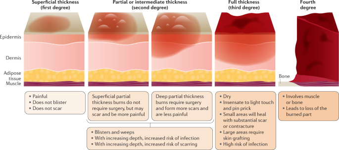
Excessive scarring is not an example of a first-degree burn. It typically occurs in more severe burns that affect deeper layers of the skin, such as second-degree or third-degree burns. Second-degree burns extend into the dermis, while third-degree burns damage all layers of the skin and can lead to significant scarring. First-degree burns, on the other hand, only affect the outer layer of the skin (epidermis) and usually do not result in excessive scarring.
B. Blistering from flames:
Blistering from flames is more characteristic of a second-degree burn rather than a first-degree burn. Second-degree burns involve damage to both the epidermis and part of the dermis, which can result in blister formation. These burns are often caused by direct contact with flames, hot liquids, or steam.
C. Blackened dead skin:
Blackened dead skin is indicative of a third-degree burn, which is the most severe type of burn. Third-degree burns damage all layers of the skin, including the epidermis, dermis, and sometimes underlying tissues. The skin may appear charred or blackened, and these burns often require medical intervention, such as skin grafting, due to the extent of tissue damage.
D. A sunburn:
A sunburn is an example of a first-degree burn. It occurs due to overexposure to ultraviolet (UV) radiation from the sun, leading to redness, pain, and mild swelling of the skin. First-degree burns affect only the outer layer of the skin (epidermis) and typically heal within a few days without significant scarring or blistering. Applying soothing lotions, staying hydrated, and avoiding further sun exposure can help manage sunburns.
Nursing Test Bank
Naxlex Comprehensive Predictor Exams
Related Questions
Correct Answer is C
Explanation
A. Cluster of oral herpes sores: Oral herpes sores typically heal within a few weeks and do not generally become chronic wounds unless there are complications or underlying immune system issues. They are more acute in nature and tend to resolve without becoming chronic.
B. Abdominal surgical incision: Surgical incisions are designed to heal within a specific timeframe, usually a few weeks to a couple of months, depending on the type of surgery and individual healing factors. While surgical wounds can sometimes have delayed healing or complications, they are not typically categorized as chronic wounds unless they fail to heal or become recurrent over an extended period.
C. Diabetic foot ulcer: Diabetic foot ulcers are highly prone to becoming chronic wounds due to the underlying pathology associated with diabetes, such as neuropathy (nerve damage), peripheral vascular disease (poor circulation), and impaired immune function. These factors can impair the normal healing process, leading to delayed healing, infection, and the potential for the wound to become chronic if not managed appropriately.
D. Posterior scalp wound: Scalp wounds can heal relatively quickly, especially with proper wound care and management. However, certain factors such as the size of the wound, depth, presence of infection, and underlying conditions can influence the likelihood of a scalp wound becoming chronic. In general, scalp wounds are less likely to become chronic compared to wounds in areas with higher risk factors, such as diabetic foot ulcers.
Correct Answer is C
Explanation
A. Increase the effectiveness of the skin graft:
Debridement can indeed increase the effectiveness of a skin graft by preparing a clean, viable wound bed for grafting. Removing dead tissue and debris helps the skin graft adhere to healthy tissue and promotes successful graft take. However, this is not the primary purpose of debridement.
B. Promote movement in the affected area:
While debridement can indirectly contribute to promoting movement by improving wound healing and reducing pain, the primary purpose of debridement is not to promote movement in the affected area.
C. Prevent infection and promote healing:
This statement accurately reflects the primary purpose of debridement. By removing nonviable tissue, debris, and foreign material from the wound, debridement helps prevent infection by reducing the bacterial load and creating an environment conducive to healing. It also promotes granulation tissue formation and wound contraction, which are essential for wound healing.
D. Promote suppuration of the wound:
Suppuration refers to the formation and discharge of pus from a wound, often indicating infection. Debridement aims to remove necrotic tissue and prevent infection, so promoting suppuration is not a desired outcome of debridement.
Whether you are a student looking to ace your exams or a practicing nurse seeking to enhance your expertise , our nursing education contents will empower you with the confidence and competence to make a difference in the lives of patients and become a respected leader in the healthcare field.
Visit Naxlex, invest in your future and unlock endless possibilities with our unparalleled nursing education contents today