A student is caring for a patient who suffered massive blood loss after trauma. How does the student correlate the blood loss with the patient's mean arterial pressure (MAP)?
It causes vasoconstriction and increased MAP.
Lower blood volume lowers MAP.
It raises cardiac output and MAP.
There is no direct correlation to MAP.
The Correct Answer is B
Mean arterial pressure (MAP) is a measure of the average pressure within the arteries during one cardiac cycle. It represents the perfusion pressure that drives blood flow to organs and tissues. MAP is calculated using the formula:
MAP = Diastolic blood pressure + 1/3 (Systolic blood pressure - Diastolic blood pressure)
Blood loss, particularly in cases of significant hemorrhage, leads to a decrease in blood volume. When blood volume decreases, there is less circulating blood available to generate pressure within the arterial system. This reduction in blood volume results in decreased MAP.
Therefore, in the case of massive blood loss after trauma, the student can correlate it with a lower blood volume, which in turn leads to a lower MAP. The decrease in blood volume reduces the perfusion pressure, compromising organ and tissue perfusion
A. It causes vasoconstriction and increased MAP in (option A) is incorrect because: While vasoconstriction can occur as a compensatory mechanism to maintain blood pressure, it does not necessarily lead to an increased MAP in the context of significant blood loss.
C. It raises cardiac output and MAP in (option C) is incorrect because Blood loss typically leads to a reduction in cardiac output due to decreased blood volume. Therefore, it does not raise cardiac output and MAP.
D. There is no direct correlation to MAP in (option D) is incorrect because: There is indeed a direct correlation between blood loss and MAP. As blood volume decreases, MAP decreases as well.
Therefore, the correct correlation between blood loss and MAP is that lower blood volume lowers MAP.
Nursing Test Bank
Naxlex Comprehensive Predictor Exams
Related Questions
Correct Answer is C
Explanation
Septic shock is a life-threatening condition characterized by severe infection, systemic inflammation, and inadequate tissue perfusion. In this critical situation, one of the initial priorities is to restore intravascular volume and improve tissue perfusion. Initiation of an intravenous line allows for the administration of fluids and other necessary medications to support the patient's hemodynamic stability.
While the other interventions mentioned are also important components of septic shock management, the immediate priority is to address hypotension and tissue hypoperfusion through fluid resuscitation:
A. Obtaining wound and blood cultures in (option A) is incorrect because: Cultures are important to identify the source and causative organisms of the infection. However, fluid resuscitation should take priority over obtaining cultures, as it is necessary to stabilize the patient's hemodynamics.
B. Removing or controlling potentially infected sources in (option B) is incorrect because: Identifying and controlling the source of infection is crucial in septic shock management to prevent further progression. However, initiating fluid resuscitation is more time-sensitive and should be prioritized.
D. Drawing blood for hematology and chemistry studies in (option D) is incorrect because Laboratory studies are important for evaluating organ function and guiding treatment. However, the immediate focus should be on fluid resuscitation to address the underlying hypoperfusion and stabilize the patient's condition.
Therefore, the intervention considered a priority when treating a patient who presents with septic shock is the initiation of an intravenous line and fluid administration to restore intravascular volume and improve tissue perfusion.
Correct Answer is A
Explanation
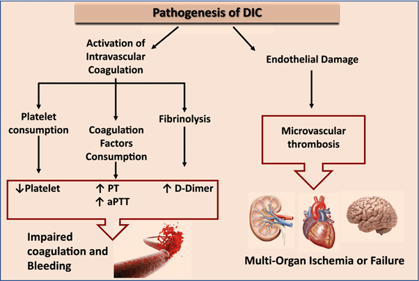
Disseminated intravascular coagulation (DIC) is a condition characterized by both widespread activation of the coagulation system and excessive clotting, leading to the consumption of clotting factors and platelets. This results in a prothrombotic state, which can lead to organ dysfunction and bleeding manifestations.
Elevated D-dimer levels are a characteristic finding in DIC. D-dimer is a fibrin degradation product that is elevated when there is excessive fibrin formation and breakdown. Elevated D-dimer indicates ongoing fibrinolysis and activation of the clotting system.
B. Decreased prothrombin time in (option B) is incorrect because: DIC is characterized by consumption of clotting factors, which can result in prolongation of the prothrombin time (PT) as well as other coagulation tests.
C. Decreased partial thromboplastin time in (option C) is incorrect because Similar to the prothrombin time, the partial thromboplastin time (PTT) can also be prolonged in DIC due to the consumption of clotting factors.
D. Elevated fibrinogen level in (option D) is incorrect because, In DIC, there is consumption of fibrinogen along with other clotting factors. Therefore, elevated fibrinogen levels are not consistent with the pathophysiology of DIC.
Whether you are a student looking to ace your exams or a practicing nurse seeking to enhance your expertise , our nursing education contents will empower you with the confidence and competence to make a difference in the lives of patients and become a respected leader in the healthcare field.
Visit Naxlex, invest in your future and unlock endless possibilities with our unparalleled nursing education contents today