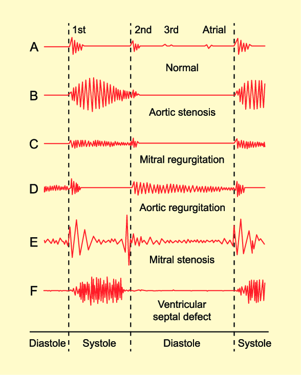
The nurse is auscultating heart sounds on a client and hears an extra sound late in diastole, just before the S1. How should the nurse document this finding?
The third heart sound (S3)
A friction rub
The fourth heart sound (S4)
A split second heart sound S2
The Correct Answer is A
A. The third heart sound (S3):
The third heart sound (S3) is an abnormal heart sound that occurs during early diastole, immediately after S2 (the second heart sound). It is caused by the rapid filling of the ventricles and is often associated with conditions like heart failure. In heart failure, the ventricles become stiff, causing vibrations that produce the S3 sound.
B. A friction rub:
A friction rub is a high-pitched, scratchy sound heard during both systole and diastole. It is caused by the rubbing together of inflamed pericardial layers (pericarditis) and is usually heard best at the left lower sternal border. Friction rubs can indicate pericardial inflammation and are often heard in conditions such as pericarditis or after a myocardial infarction.
C. The fourth heart sound (S4):
The fourth heart sound (S4) occurs late in diastole, just before S1, and is caused by atrial contraction. It is associated with increased resistance to ventricular filling, often due to conditions like hypertension or aortic stenosis. The S4 sound is heard as a low-pitched "atrial gallop."
D. A split second heart sound S2:
The second heart sound (S2) represents the closure of the aortic and pulmonic valves. Normally, S2 has two components: A2 (aortic valve closure) and P2 (pulmonic valve closure). A split S2 occurs when A2 and P2 do not close simultaneously. A physiological split S2 is common during inspiration and occurs due to delayed closure of the pulmonic valve. An abnormal or fixed split S2 can indicate underlying heart conditions such as atrial septal defect (ASD) or right bundle branch block (RBBB).
Nursing Test Bank
Naxlex Comprehensive Predictor Exams
Related Questions
Correct Answer is D
Explanation
. Presence of breath sounds: While assessing the anterior chest, the nurse should listen for breath sounds over various areas of the lungs. However, this is related to auscultation, not inspection.
B. Diaphragmatic excursion: Diaphragmatic excursion involves assessing the movement of the diaphragm during breathing. This is typically done by percussing the level where dullness changes to resonance during inhalation and exhalation. It is more related to percussion, not inspection.
C. Symmetric chest expansion: Symmetric chest expansion refers to the equal expansion of both sides of the chest during inhalation. The nurse can observe and palpate the chest to assess if it expands symmetrically on both sides. This is a crucial aspect of the inspection of the anterior chest.
D. Shape and configuration of the chest wall: The shape and configuration of the chest wall, including abnormalities or deformities, should be assessed during inspection. This includes observing for any asymmetry, deformities, masses, or scars on the anterior chest.
Correct Answer is D
Explanation
A. When bronchial breath sounds are auscultated in the trachea.
Auscultating bronchial breath sounds in the trachea is a normal finding, as the trachea is close to the upper airway, and this is where bronchial sounds are normally heard. However, if these sounds are heard in the peripheral lung fields, it can indicate an abnormal condition.
B. When the client is experiencing excessive sneezing from a tree pollen allergy.
Excessive sneezing due to allergies would not typically result in increased breath sounds. Allergies may cause nasal congestion, but they don't directly lead to increased breath sounds.
C. When the client is resting in bed and not experiencing respiratory issues.
If a client is at rest and not experiencing any respiratory issues, breath sounds should typically be normal. There would be no reason to expect increased breath sounds in this scenario.
D. When the bronchial tree is obstructed by secretions.
Increased breath sounds, such as wheezing or rhonchi, can be auscultated when there is an obstruction in the bronchial tree due to secretions, narrowing of the airways, or other causes. These sounds are typically abnormal and indicate an issue with air movement through the airways.
Whether you are a student looking to ace your exams or a practicing nurse seeking to enhance your expertise , our nursing education contents will empower you with the confidence and competence to make a difference in the lives of patients and become a respected leader in the healthcare field.
Visit Naxlex, invest in your future and unlock endless possibilities with our unparalleled nursing education contents today