When the nurse checks the patient for orthodontic hypotension, what did the nurse have the patient do?
Physical exertion
Eat
Stand up
D. Lie down
The Correct Answer is C
Orthostatic hypotension is a drop in blood pressure that occurs when a person stands up from a sitting or lying down position. To check for orthostatic hypotension, the nurse typically takes the patient's blood pressure and heart rate while the patient is lying down, then has the patient stand up for a few minutes and takes the blood pressure and heart rate again. If the blood pressure drops significantly (usually a drop of 20 mm Hg or more) and the heart rate increases, it may indicate orthostatic hypotension.
Nursing Test Bank
Naxlex Comprehensive Predictor Exams
Related Questions
Correct Answer is C
Explanation
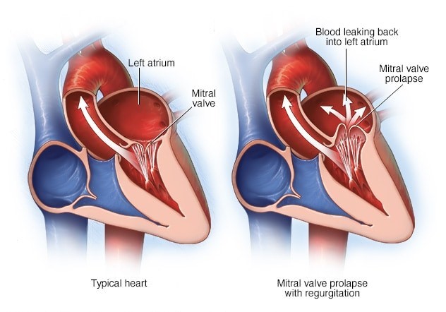
Mitral stenosis refers to a narrowing of the mitral valve, which is located between the left atrium and the left ventricle. This narrowing can cause incomplete emptying of blood from the left atrium into the left ventricle during diastole (relaxation phase) of the cardiac cycle. This can lead to increased pressure in the left atrium and pulmonary circulation, causing symptoms such as shortness of breath, fatigue, and pulmonary con
Correct Answer is D
Explanation
Sustained hypertension can lead to several complications, including damage to the blood vessels in the retina (retinopathy), increased risk of stroke, and damage to the kidneys (renal disease). Other potential complications include heart disease, peripheral arterial disease, and cognitive impairment. It is important to manage hypertension through lifestyle modifications and medication to prevent these complications.
Whether you are a student looking to ace your exams or a practicing nurse seeking to enhance your expertise , our nursing education contents will empower you with the confidence and competence to make a difference in the lives of patients and become a respected leader in the healthcare field.
Visit Naxlex, invest in your future and unlock endless possibilities with our unparalleled nursing education contents today