
Anatomy and physiology II
Anatomy and physiology II
Total Questions : 39
Showing 10 questions Sign up for moreWhich of the following cartilages is largest?
Explanation
A. Corniculate cartilage: Small horn-shaped cartilages that sit on top of the arytenoid cartilages; not the largest.
B. Epiglottic cartilage: Leaf-shaped cartilage that covers the glottis during swallowing; important but smaller than the thyroid cartilage.
C. Thyroid cartilage: This is the largest laryngeal cartilage; it forms the laryngeal prominence (Adam’s apple).
D. Cricoid cartilage: Ring-shaped and located below the thyroid cartilage; it is smaller than the thyroid cartilage but thicker posteriorly.
E. Arytenoid cartilage: Pyramid-shaped cartilages involved in vocal cord movement; smaller in size.
Explanation
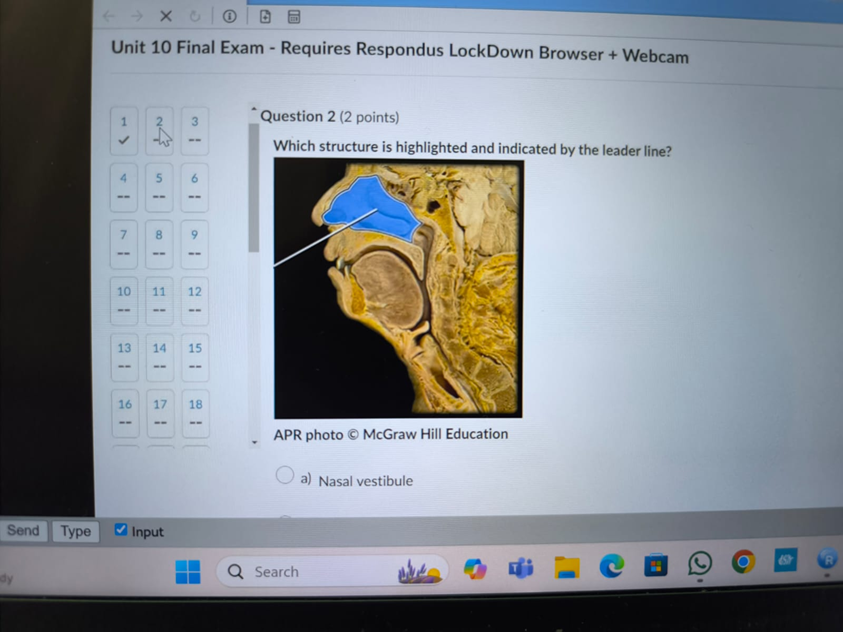
A. Nasal vestibule: The nasal vestibule is the anterior part of the nasal cavity just inside the nostrils, lined with skin and hair.
B. Nasal cavity: The nasal cavity is a large air-filled space behind the nose, extending from the nostrils to the choanae.
C. Nasal concha: The nasal conchae are curved bony projections (superior, middle, and inferior) inside the nasal cavity that increase the surface area for warming and humidifying air.
D. Sphenoid sinus: The sphenoid sinus is a hollow space within the sphenoid bone, located posterior to the nasal cavity and inferior to the sella turcica.
Which of the following would lead to anemic hypoxia?
Explanation
A. Sickle-cell disease: Anemic hypoxia occurs when the oxygen-carrying capacity of the blood is reduced, as in sickle-cell disease where abnormal hemoglobin impairs oxygen transport.
B. Emphysema: This causes hypoxic hypoxia due to impaired gas exchange, not anemic hypoxia.
C. Squamous-cell carcinoma: May obstruct airways but does not directly cause anemic hypoxia.
D. Asthma: Causes obstructive hypoxia (ventilatory defect), not a reduction in hemoglobin or oxygen-carrying capacity.
E. Atelectasis: Collapse of alveoli reduces oxygenation but does not reduce the hemoglobin content-this is hypoxic hypoxia.
Hypocapnia will lead to which of the following conditions?
Explanation
A. Hypoventilation due to acidosis: Hypoventilation would increase CO₂, leading to respiratory acidosis, not hypocapnia.
B. Hypoventilation due to alkalosis: Hypocapnia is a result of hyperventilation, not hypoventilation.
C. Hyperventilation due to acidosis: Hyperventilation is a response to acidosis to blow off CO₂, but hypocapnia itself is a result of hyperventilation.
D. Hyperventilation due to alkalosis: Hypocapnia (low CO₂) leads to respiratory alkalosis. This occurs due to hyperventilation, which blows off too much CO₂.
E. Hypocapnia does not affect ventilation rate: CO₂ levels significantly influence ventilation via central chemoreceptors.
The lungs have a total of five
Explanation
A. Segmental bronchi: There are more than five segmental (tertiary) bronchi; each lung has multiple segments.
B. Choanae: These are posterior nasal apertures, not part of the lungs.
C. Laryngeal cartilages: There are nine laryngeal cartilages in total, not five in the lungs.
D. Lobes: The right lung has three lobes (superior, middle, inferior), and the left lung has two lobes-totaling five.
E. Tracheal cartilages: The trachea has multiple C-shaped cartilaginous rings, not five.
Explanation
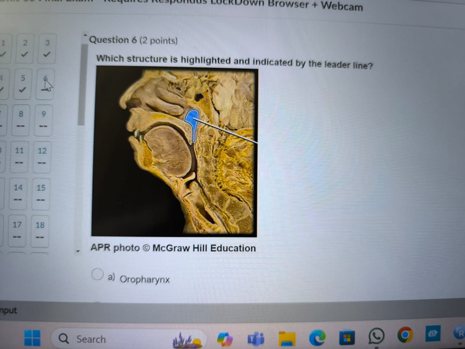
A. Oropharynx: The oropharynx is the middle section of the pharynx, located posterior to the oral cavity and extending from the soft palate to the epiglottis.
B. Nasopharynx: The nasopharynx is the uppermost part of the pharynx, located posterior to the nasal cavity and above the soft palate. The highlighted structure in the image lies just behind the nasal cavity and in front of the cervical spine, matching the location of the nasopharynx.
C. Hypopharynx: The hypopharynx (laryngopharynx) is the lower part of the pharynx, located below the oropharynx and above the esophagus, behind the larynx.
D. Esophagus: The esophagus is the muscular tube that carries food from the pharynx to the stomach. It lies inferior and posterior to the larynx and trachea.
Explanation
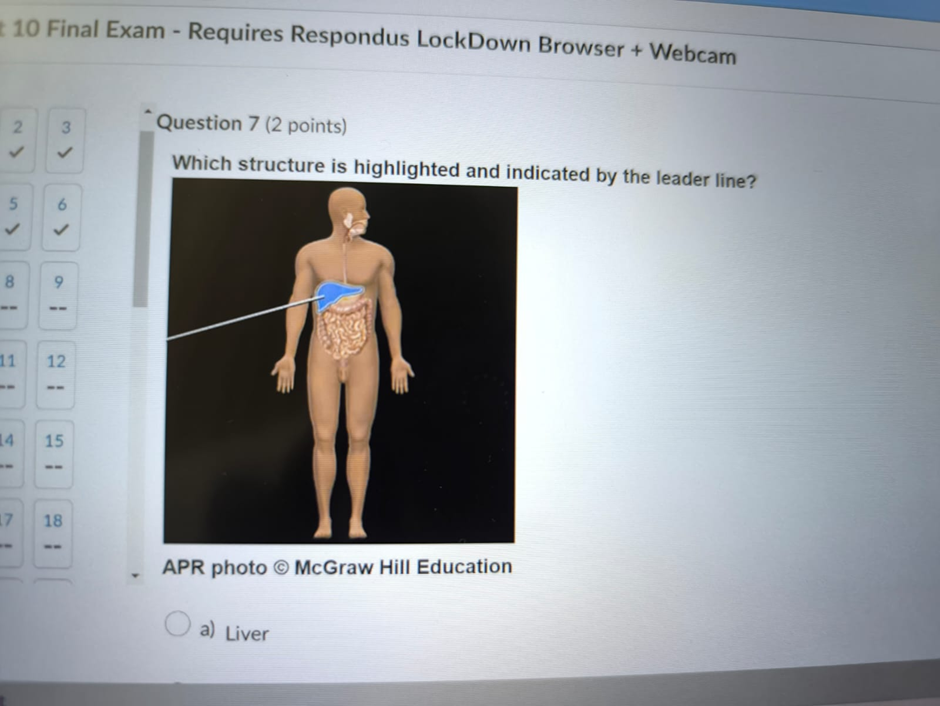
A. Liver: The liver is a large, reddish-brown organ in the right upper quadrant of the abdomen, just beneath the diaphragm. If the diagram is pointing to this area, especially the largest solid organ on the right side, it matches the liver’s location and size.
B. Stomach: The stomach is located mainly in the left upper quadrant, inferior to the diaphragm and to the left of the liver. It is smaller and has a J-shaped structure, so it would not match if the pointer is on the large right-sided organ.
C. Spleen: The spleen is located in the left upper quadrant, lateral to the stomach and posterior to the rib cage. It is much smaller than the liver and not in the right-sided position indicated.
D. Diaphragm: The diaphragm is a dome-shaped muscle that separates the thoracic and abdominal cavities. If the pointer is on the muscle just beneath the lungs, it would be the diaphragm, but here it’s on a large organ, not a muscle.
Explanation
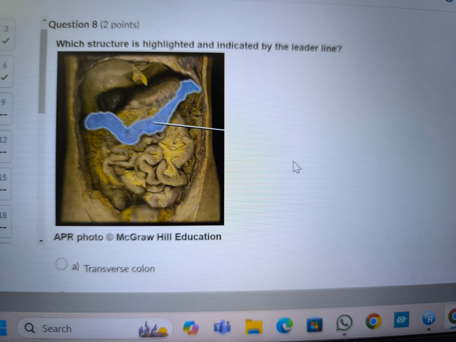
A. Transverse colon: The transverse colon is the horizontal portion of the large intestine that runs across the upper abdomen from the right colic flexure (near the liver) to the left colic flexure (near the spleen).
B. Ascending colon: The ascending colon runs vertically upward along the right side of the abdomen, from the cecum to the hepatic flexure.
C. Descending colon: The descending colon runs vertically down the left side of the abdomen, from the splenic flexure to the sigmoid colon.
D. Ileum: The ileum is the final section of the small intestine, located in the lower abdomen, and connects to the cecum.
Explanation
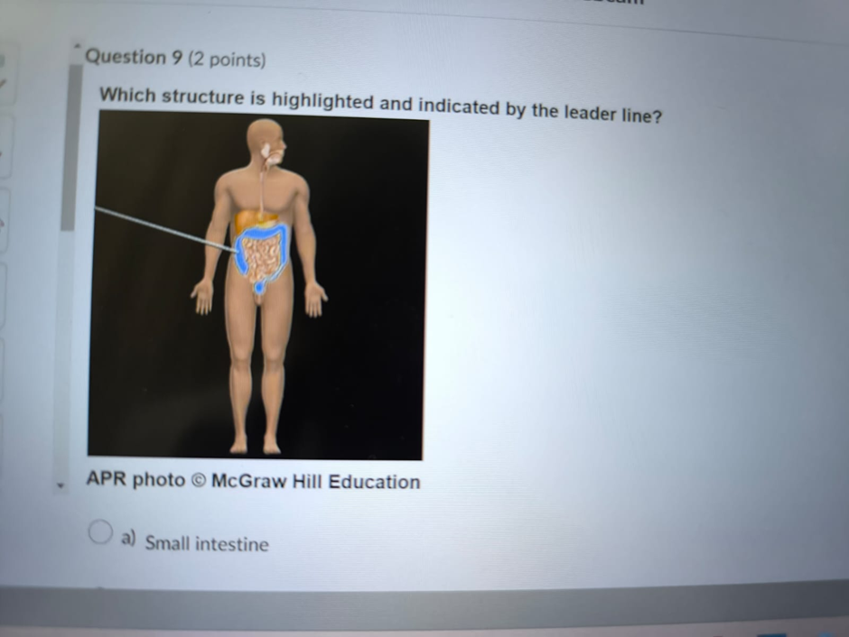
A. Small intestine: The small intestine is a long, coiled tube occupying most of the central and lower abdomen, made up of the duodenum, jejunum, and ileum.
B. Large intestine: The large intestine frames the small intestine and includes the cecum, colon, rectum, and anal canal. It is wider in diameter and often shown outlining the abdominal cavity.
C. Rectum: The rectum is the terminal portion of the large intestine, located in the pelvic cavity, and does not extend throughout the abdomen.
D. Caecum: The cecum is the first part of the large intestine, located in the right lower quadrant, just below the ileocecal valve.
The
Explanation
A. Gastric rugae: Ridges/folds in the stomach lining that allow it to expand, but do not regulate flow into the duodenum.
B. Antrum: The lower portion of the stomach that grinds food and mixes it with gastric juices; helps move chyme but does not regulate its release.
C. Pyloric sphincter: A muscular valve at the distal end of the stomach that controls the passage of chyme into the duodenum.
D. Fundus: The upper rounded portion of the stomach; involved in food storage, not in controlling outflow.
E. Cardial part: Connects the esophagus to the stomach and receives food; not involved in regulating outflow to the duodenum.
You just viewed 10 questions out of the 39 questions on the Anatomy and physiology II Exam. Subscribe to our Premium Package to obtain access on all the questions and have unlimited access on all Exams. Subscribe Now

