
Please set your exam date
Disorders of the Eye
Study Questions
Practice Exercise 1
What is in the posterior cavity of the eye?
Explanation
The posterior cavity of the eye, located behind the lens and in front of the retina, is filled with vitreous humor. This gel-like substance helps maintain the eye’s shape, provides structural support, and keeps the retina in place by pressing it against the choroid.
Rationale for correct answer:
4. Vitreous humor: This transparent, jelly-like fluid fills the posterior cavity. It plays a crucial role in maintaining intraocular pressure, supporting the lens, and ensuring that light is properly focused onto the retina.
Rationale for incorrect answers:
- Zonules: These are suspensory ligaments that attach the lens to the ciliary body. They are not located in the posterior cavity but in the area around the lens.
- Cornea: The cornea is part of the anterior segment, forming the transparent dome-shaped surface at the front of the eye.
- Aqueous humor: This watery fluid fills the anterior and posterior chambers of the anterior segment, not the posterior cavity.
Take home points
- The posterior cavity lies between the lens and the retina.
- It contains vitreous humor, not aqueous humor.
- Structures like the cornea and zonules belong to the anterior segment of the eye.
What is the function of the sclera?
Explanation
The sclera is the protective white outer layer of the eyeball. It provides structural support, helps maintain the shape of the eye, and serves as an attachment site for extraocular muscles. This tough, fibrous layer shields the inner components of the eye from injury and damage.
Rationale for correct answer:
3. Protective white outer layer of the eyeball: The sclera acts as a tough outer coat that maintains the shape of the eye and protects delicate internal structures while serving as a point of attachment for the ocular muscles.
Rationale for incorrect answers:
1. Secrete aqueous humor: This function is performed by the ciliary body, not the sclera.
2. Focus light rays on the retina: This is the role of the cornea and lens, not the sclera.
4. Photoreceptor cells stimulated in dim environments: These are rods in the retina, not part of the sclera.
Take home points
- The sclera is the tough, white, protective outer layer of the eye.
- It does not secrete aqueous humor, focus light, or detect stimuli.
- It provides protection, shape, and muscle attachment for the eyeball.
The nurse documents PERRLA following assessment of a patient’s eyes. What is one finding that supports this statement?
Explanation
PERRLA stands for Pupils Equal, Round, Reactive to Light, and Accommodation. A normal finding that supports this assessment is constriction of the pupils when an object is brought closer to the eyes, which demonstrates accommodation. This reflex ensures that the eyes can focus on near objects effectively.
Rationale for correct answer:
4. Constriction of the pupils when an object is brought closer to the eyes: This confirms accommodation, which is part of the PERRLA assessment. It ensures the pupils respond appropriately to focusing demands.
Rationale for incorrect answers:
- A slightly oval shape of the pupils: Pupils should be round in a normal PERRLA finding. An oval pupil may indicate pathology.
- The presence of nystagmus on far lateral gaze: Nystagmus is an involuntary eye movement and is not part of a normal PERRLA assessment.
- Dilation of the pupil when a light is shined in the opposite eye: This is the opposite of what should occur. Normally, the opposite pupil should also constrict (consensual response).
Take home points
- PERRLA = Pupils Equal, Round, Reactive to Light, and Accommodation.
- A key confirming sign is pupil constriction with accommodation.
- Abnormal findings like oval pupils, nystagmus, or paradoxical dilation do not support PERRLA.
To determine the presence of corneal abrasions or defects in a patient with an eye injury, what would the nurse provide?
Explanation
The nurse uses fluorescein dye to detect corneal abrasions or defects. When applied to the eye, the dye adheres to damaged areas of the cornea, which then appear bright green under a blue light (Wood’s lamp or slit lamp). This method is quick, noninvasive, and highly effective for identifying even small injuries.
Rationale for correct answer:
2. Fluorescein dye: This dye highlights corneal defects by staining epithelial breaks. Under blue light, the damaged area glows, confirming abrasions or ulcers.
Rationale for incorrect answers:
1. Tonometer: This instrument measures intraocular pressure, typically used to assess for glaucoma, not corneal injury.
3. Pocket penlight: A penlight is useful for assessing pupil reactions but cannot detect corneal abrasions.
4. An ophthalmoscope: This tool is designed for examining the retina and internal eye structures, not for corneal surface injuries.
Take home points
- Fluorescein dye is the standard tool to detect corneal abrasions or epithelial defects.
- Tonometer → intraocular pressure, Penlight → pupils, Ophthalmoscope → retina.
- Always use blue light after fluorescein instillation to visualize abrasions.
What are possible abnormal assessment findings when assessing the eyelid? Select all that apply
Explanation
When assessing the eyelid, the nurse may encounter abnormal findings such as ptosis and blepharitis. Ptosis refers to drooping of the upper eyelid, while blepharitis is inflammation of the eyelid margins. Both conditions are directly related to eyelid abnormalities.
Rationale for correct answers:
1. Ptosis: Drooping of the upper eyelid due to neuromuscular disorders (e.g., myasthenia gravis) or cranial nerve III dysfunction.
3. Blepharitis: Inflammation of the eyelid margins, often caused by infection, seborrheic dermatitis, or dysfunction of meibomian glands.
Rationale for incorrect answers:
2. Strabismus: Misalignment of the eyes, not an eyelid abnormality.
4. Anisocoria: Unequal pupil size, an abnormality of the pupils, not the eyelid.
5. Swelling of the pinna: Refers to the external ear, not related to eyelid assessment.
Take home points
- Abnormal eyelid findings include ptosis (drooping) and blepharitis (inflammation).
- Strabismus and anisocoria involve the eyes and pupils, not the eyelid.
- Swelling of the pinna is an ear finding, not an eyelid abnormality.
Practice Exercise 2
A patient with early cataracts tells the nurse that he is afraid cataract surgery may cause permanent visual damage. What should the nurse teach the patient?
Explanation
For a patient with early cataracts, the nurse should emphasize that vision enhancement techniques can help improve quality of life until surgery is needed. These include stronger prescription glasses, use of magnifiers, and increased lighting. Cataract surgery is generally safe and effective, but it is typically postponed until the cataracts significantly interfere with daily activities.
Rationale for correct answer:
4. Vision enhancement techniques may improve vision until surgery becomes an acceptable option to maintain desired activities: This teaching reassures the patient and provides practical measures to cope with vision changes until surgery is appropriate.
Rationale for incorrect answers:
- The cataracts will only worsen with time and should be removed as early as possible to prevent blindness: Cataracts do progress, but they do not require immediate removal unless they impair daily function. Early removal is not always necessary.
- Cataract surgery is very safe and with the implantation of an intraocular lens, the need for glasses will be eliminated: Surgery is safe, but many patients still need glasses postoperatively for fine vision tasks.
- Progression of the cataracts can be prevented by avoidance of ultraviolet (UV) light and good dietary management: These measures may reduce risk but cannot stop or reverse cataract progression once it has begun.
Take home points
- In early cataracts, surgery is not immediately required.
- Vision enhancement techniques can maintain independence until surgery becomes necessary.
- Cataracts cannot be prevented or reversed by lifestyle measures once present, and surgery may still require corrective lenses afterward.
A 60-year-old patient is being prepared for outpatient cataract surgery. When obtaining admission data from the patient, what would the nurse expect to find in the patient’s history?
Explanation
In cataracts, the typical history is gradual loss of vision, accompanied by abnormal color perception and glare sensitivity. This results from clouding of the eye’s natural lens, which scatters incoming light and impairs sharp visual focus. Unlike other eye conditions, cataracts develop slowly and painlessly.
Rationale for correct answer:
3. A gradual loss of vision with abnormal color perception and glare: This is the hallmark of cataracts. Patients often describe cloudy or dim vision, fading of colors, and difficulty with glare, especially at night.
Rationale for incorrect answers:
1. A painless, sudden, severe loss of vision: This suggests retinal artery occlusion or another acute event, not cataracts.
2. Blurred vision, colored halos around lights, and eye pain: These are typical of acute angle-closure glaucoma, not cataracts.
4. Light flashes, floaters, and a “cobweb” in the field of vision with loss of central or peripheral vision: These are signs of retinal detachment, not cataracts.
Take home points
- Cataracts cause gradual, painless vision loss with color distortion and glare.
- Acute vision loss, halos with pain, or floaters/flashes suggest other serious eye conditions, not cataracts.
- Careful history helps differentiate cataracts from glaucoma and retinal disorders.
Which characteristics of glaucoma are associated with only primary open-angle glaucoma (POAG)? Select all that apply
Explanation
Primary open-angle glaucoma (POAG) is the most common type of glaucoma. It is characterized by a gradual, painless loss of peripheral vision due to resistance to aqueous outflow through the trabecular meshwork. POAG is treated medically, often with cholinergic agents such as pilocarpine, which increase aqueous humor outflow.
Rationale for correct answers:
1. Gradual loss of peripheral vision: This is a classic sign of POAG, progressing slowly and often unnoticed until advanced.
5. Treated with cholinergic agents such as pilocarpine (Pilocar): Medications are the mainstay of POAG treatment, aiming to reduce intraocular pressure.
6. Resistance to aqueous outflow through trabecular meshwork: This is the underlying pathophysiology of POAG.
Rationale for incorrect answers:
2. Treated with iridotomy or iridectomy: This surgical procedure is used in angle-closure glaucoma, not POAG.
3. Causes loss of central vision with corneal edema: These are features of acute angle-closure glaucoma, which presents suddenly and painfully.
4. May be caused by increased production of aqueous humor: Overproduction is not the usual cause of POAG; the problem lies with impaired drainage.
Take home points
- POAG → gradual peripheral vision loss, resistance at trabecular meshwork, treated with medications (e.g., pilocarpine).
- Angle-closure glaucoma → sudden painful vision loss, corneal edema, treated with iridotomy/iridectomy.
- Differentiating POAG from angle-closure is essential for correct management.
For the patient with a retinal break, what extraocular techniques may be used with sclera buckling to seal the break by creating an inflammatory reaction that causes a chorioretinal adhesion or scar? Select all that apply
Explanation
With scleral buckling, extraocular techniques such as cryopexy and laser photocoagulation are used to create a controlled inflammatory response that produces chorioretinal adhesion or scar formation. This seals the retinal break and prevents further detachment.
Rationale for correct answers:
1. Cryopexy: A freezing probe applied externally over the sclera induces inflammation and scar tissue formation around the retinal break.
4. Laser photocoagulation: A laser beam creates small burns around the retinal tear, leading to scar tissue formation that seals the break.
Rationale for incorrect answers:
2. Vitrectomy: This is an intraocular procedure that removes the vitreous humor to relieve traction on the retina; it is not an extraocular technique.
3. Pneumatic retinopexy: Involves injection of a gas bubble inside the vitreous cavity; it is also an intraocular method, not extraocular.
5. Penetrating keratoplasty: This is a corneal transplant procedure, unrelated to retinal repair.
Take home points
- Extraocular sealing techniques with scleral buckling include cryopexy (freezing) and laser photocoagulation (burning).
- Vitrectomy and pneumatic retinopexy are intraocular methods.
- Penetrating keratoplasty is a corneal procedure, not related to retinal breaks.
What nursing action is most important for the patient with age-related macular degeneration (AMD)?
Explanation
For patients with age-related macular degeneration (AMD), the most important nursing intervention is to emphasize vision enhancement techniques. These strategies help patients maximize remaining vision and maintain independence. They may include using magnifiers, special lenses, bright lighting, and visual aids. While certain treatments exist for the wet form of AMD (e.g., anti-VEGF injections), vision cannot be fully restored, so supportive measures are essential.
Rationale for correct answer:
2. Emphasize the use of vision enhancement techniques to improve what vision is present: These measures enable patients to adapt to central vision loss and maintain daily functioning despite irreversible damage.
Rationale for incorrect answers:
1. Teach the patient how to use topical eyedrops for treatment of AMD: No effective topical eyedrops exist for AMD treatment.
3. Encourage the patient to undergo laser treatment to slow the deposit of extracellular debris: Laser therapy is not typically used; wet AMD is managed with intravitreal injections, not laser for drusen deposits.
4. Explain that nothing can be done to save the patient’s vision because there is no treatment for AMD: This is incorrect and discouraging. While AMD cannot be cured, supportive interventions and some treatments for wet AMD can help.
Take home points
- AMD leads to central vision loss, but supportive strategies help patients adapt.
- Vision enhancement techniques (magnifiers, lighting, low-vision aids) are critical nursing interventions.
- There is no cure, but wet AMD can be managed with anti-VEGF therapy.
Practice Exercise 3
A patient with wet AMD is treated with photodynamic therapy. What does the nurse instruct the patient to do after the procedure?
Explanation
After photodynamic therapy for wet AMD, the nurse must stress strict avoidance of direct sunlight or bright indoor light for several days. The photosensitizing drug used in the procedure remains active in the body and can cause a thermal burn if exposed to sunlight. Patients are instructed to completely cover their skin and eyes when outside.
Rationale for correct answer:
3. Completely cover all the skin to avoid a thermal burn from sunlight: The photosensitizing drug (verteporfin) can be activated by sunlight, leading to burns. Protection is critical after the procedure.
Rationale for incorrect answers:
1. Maintain the head in an upright position for 24 hours: This instruction applies to pneumatic retinopexy, not photodynamic therapy.
2. Avoid blowing the nose or causing jerking movements of the head: These precautions are associated with retinal surgery, not this therapy.
4. Expect to experience blind spots where the laser has caused retinal damage: Photodynamic therapy is designed to minimize retinal damage; blind spots are not expected.
Take home points
- After photodynamic therapy for wet AMD, patients must avoid sunlight/bright light exposure for several days.
- Teach patients to cover skin and eyes completely to prevent burns.
- Precautions differ from those for retinal detachment surgery or laser therapy.
What is an important health promotion nursing intervention related to glaucoma?
Explanation
The most important health promotion intervention for glaucoma is regular intraocular pressure (IOP) screening. Since glaucoma often progresses silently without early symptoms, routine eye exams are vital for early detection and treatment, which can prevent irreversible optic nerve damage and vision loss.
Rationale for correct answer:
3. Promoting regular measurements of intraocular pressure for early detection and treatment of glaucoma: Screening detects glaucoma before symptoms appear, allowing treatment to slow progression and preserve vision.
Rationale for incorrect answers:
1. Teaching individuals at risk for glaucoma about early signs and symptoms of the disease: Glaucoma is often asymptomatic until advanced, so relying on symptom recognition is ineffective.
2. Preparing patients with glaucoma for lifestyle changes necessary to adapt to eventual blindness: This is unnecessarily pessimistic; many patients retain useful vision with treatment.
4. Informing patients that glaucoma is curable if eye medications are administered before visual impairment has occurred: Glaucoma is not curable; it is a chronic disease managed with medications or surgery.
Take home points
- Glaucoma health promotion = regular IOP screenings for early detection.
- It is a chronic, controllable disease but not curable.
- Relying on symptom recognition is ineffective because glaucoma is often silent until late stages.
A patient with bilateral cataracts is scheduled for an extracapsular cataract extraction with an intraocular lens implantation of one eye. What should be done by the nurse preoperatively?
Explanation
Before cataract surgery, the nurse should assess the visual acuity of the unoperated eye. This helps determine how much assistance the patient will need postoperatively, since vision in the operated eye may be blurry initially during healing. Proper planning ensures patient safety and independence after surgery.
Rationale for correct answer:
1. Assess the visual acuity in the unoperated eye to plan the need for postoperative assistance: This is a key preoperative step to prepare for postoperative adaptation and safety needs.
Rationale for incorrect answers:
2. Inform the patient that the operative eye will need to be patched for 3 to 4 days postoperatively: Eye patching is usually only for 24 hours or may not be required at all with modern techniques.
3. Assure the patient that vision in the operative eye will be improved to near normal on the first postoperative day: Vision improvement occurs gradually, not immediately.
4. Teach the patient routine coughing and deep-breathing techniques to use postoperatively to prevent respiratory complications: This is important after many surgeries, but after eye surgery, coughing should actually be minimized to prevent increased intraocular pressure.
Take home points
- Pre-op cataract care: Assess visual acuity in the unoperated eye to plan assistance.
- Vision is not instantly restored after surgery.
- Coughing/straining should be avoided post-op to protect the operative eye.
Following a pneumatic retinopexy, what does the nurse need to know about the postoperative care for the patient?
Explanation
After a pneumatic retinopexy, the patient must follow strict positioning and activity restrictions for several days. This positioning allows the intraocular gas bubble to press against the retinal break, facilitating healing and reattachment. Adherence to these instructions is critical for the success of the procedure.
Rationale for correct answer:
1. Specific positioning and activity restrictions are likely to be required for several days: This is essential to ensure the gas bubble remains in contact with the retinal break and promotes reattachment.
Rationale for incorrect answers:
2. The patient is frequently hospitalized for 7 to 10 days on bed rest until healing is complete: Pneumatic retinopexy is an outpatient procedure, not one requiring prolonged hospitalization.
3. Patients experience little or no pain, and development of pain indicates hemorrhage or infection: Pain is not a typical feature, but the key postoperative concern is positioning, not pain.
4. Reattachment of the retina commonly fails, and patients can be expected to grieve for loss of vision: Retinal reattachment is often successful with proper care, so this statement is inaccurate and unnecessarily discouraging.
Take home points
- Strict positioning/activity restriction is required post-op to keep the gas bubble in place.
- Hospitalization is not usually required as this is an outpatient treatment.
- Pain is not the main concern; focus is on adherence to positioning.
- Retinal reattachment is usually successful if post-op instructions are followed.
What advice would the nurse give to a client who has just undergone fluorescein angiography? Select all that apply
Explanation
After fluorescein angiography, it is normal for the patient’s skin to appear slightly yellow and for the urine to be bright yellow for a short duration (up to 24–36 hours). These changes result from the excretion of fluorescein dye through the kidneys.
Rationale for correct answers:
4. Expect skin to appear slightly yellow for 6 to 8 hours: The dye temporarily causes yellow discoloration of the skin.
5. Expect urine to appear bright yellow for 24 to 36 hours: The dye is excreted renally, resulting in bright yellow urine.
Rationale for incorrect answers:
- Expect hives or rashes within 4 hours: Hives and rashes are abnormal and indicate an allergic reaction, not a normal effect.
- Expect mild headaches for 24 to 36 hours: Headaches are not a typical side effect.
- Expect red and swollen eyes for 6 to 9 hours: The test involves dye injection, not direct trauma to the eyes, so this is not expected.
Take home points
- Normal effects: Temporary yellow skin and bright yellow urine.
- Abnormal effects: Rash, hives, or breathing difficulty = possible allergic reaction → report immediately.
- The dye clears naturally within 24–36 hours.
Comprehensive Questions
A nurse is caring for an older adult client who has diabetes mellitus. The client reports loss of peripheral vision. For which of the following is the client at risk?
Explanation
Loss of peripheral vision is a classic symptom of open-angle glaucoma, a condition caused by gradual damage to the optic nerve. This occurs when aqueous humor does not drain effectively, leading to increased intraocular pressure. The condition develops slowly, and many clients are unaware until vision changes become significant. Older adults and individuals with diabetes are at higher risk of developing open-angle glaucoma.
Rationale for correct answer:
2. Open-angle glaucoma: Causes a gradual, painless loss of peripheral vision that progresses to tunnel vision if untreated. It is the most common type of glaucoma and is strongly associated with aging and diabetes.
Rationale for incorrect answers:
1. Cataracts: Cause blurred or cloudy vision and sensitivity to glare but do not affect peripheral vision. The vision loss is more generalized.
3. Macular degeneration: Leads to central vision loss, making it hard to read or recognize faces, but peripheral vision remains intact.
4. Angle-closure glaucoma: Presents suddenly with eye pain, halos, headache, and nausea, not gradual peripheral vision loss.
Take home points
- Peripheral vision loss → Open-angle glaucoma.
- Central vision loss → Macular degeneration.
- General cloudy vision → Cataracts.
- Sudden painful vision loss → Angle-closure glaucoma.
A nurse is caring for a client following a trabeculectomy. Which of the following statements should the nurse include in the teaching?
Explanation
After a trabeculectomy, the client must avoid activities that increase intraocular pressure, such as bending, lifting, or straining, to allow proper healing of the surgical site. Engaging in strenuous housekeeping tasks may put pressure on the eye and compromise surgical success. Therefore, limiting housekeeping activities is a crucial part of postoperative teaching to promote recovery and prevent complications. Education focuses on lifestyle adjustments that protect the surgical site and vision during the healing phase.
Rationale for correct answer:
4. You need to limit your housekeeping activities: Housekeeping tasks often involve bending, lifting, and straining, which can increase intraocular pressure and compromise the healing of the surgical site. Teaching the patient to limit these activities promotes proper healing and reduces the risk of postoperative complications.
Rationale for incorrect answers:
- You may resume playing golf: Golf requires significant bending and exertion, which may elevate intraocular pressure and endanger healing after trabeculectomy. It is not safe until cleared by the ophthalmologist.
- You need to tilt your head back when washing your hair: Tilting the head back can cause water or shampoo to enter the eye, increasing the risk of infection or irritation. This practice is not recommended for patients after eye surgery.
- You may continue driving to and from work: Driving may not be safe immediately after trabeculectomy due to potential blurred vision, discomfort, or risk of increased eye strain. It is unsafe until the patient is cleared by their eye specialist.
Take home points
- Patients must limit strenuous activities such as housekeeping after trabeculectomy.
- Golf, driving, and improper head positioning when washing hair are not safe until cleared by the provider.
- Protecting the eye from strain and pressure is essential for healing and preventing complications.
A nurse is caring for a male older adult client who has a new diagnosis of glaucoma. Which of the following should the nurse recognize as risk factors associated with this disease? Select all that apply
Explanation
Glaucoma is a progressive optic neuropathy often associated with increased intraocular pressure, leading to vision loss if untreated. Several risk factors increase the likelihood of disease, including age, family history, diabetes, and hypertension. Early identification of these risk factors allows for screening and early management, which is crucial in preventing irreversible blindness. The nurse should recognize and educate patients about both modifiable and non-modifiable risk factors to promote early intervention.
Rationale for correct answers:
2. Genetic predisposition: A family history of glaucoma significantly increases the client’s likelihood of developing the disease. Genetics is a recognized risk factor for glaucoma.
3. Hypertension: High blood pressure can impair blood flow to the optic nerve, increasing the risk of glaucomatous damage. Hypertension is a recognized risk factor for glaucoma.
4. Age: Advancing age is one of the strongest risk factors for glaucoma, with prevalence increasing significantly after age 60. Age is a recognized risk factor for glaucoma.
5. Diabetes mellitus: Diabetes contributes to microvascular damage, which increases susceptibility to optic nerve damage and glaucoma. Diabetes is a recognized risk factor for glaucoma.
Rationale for incorrect answer:
1. Gender: Gender alone is not considered a significant risk factor for the development of glaucoma. Both men and women are affected, and risk is more closely related to age and systemic conditions.
Take home points
- Major risk factors for glaucoma include genetics, hypertension, advanced age, and diabetes.
- Gender is not a direct risk factor for glaucoma.
- Nurses play a critical role in identifying at-risk patients and encouraging regular eye exams for early detection.
A nurse is caring for a client who has a new diagnosis of cataracts. Which of the following clinical manifestations should the nurse expect to find? Select all that apply
Explanation
Cataracts are a progressive eye condition caused by the clouding of the lens, leading to a gradual decrease in vision. Common clinical manifestations include blurred vision, difficulty with glare, and the appearance of a white pupil or lens opacity. Since the onset is gradual, patients may not notice early changes until the cataract progresses. Recognizing these hallmark manifestations is important for timely intervention and patient education.
Rationale for correct answers:
3. Blurred vision: Cataracts interfere with light transmission through the lens, leading to cloudy or blurred vision. Blurred vision is a recognized clinical manifestation of cataracts.
4. White pupils: As cataracts progress, the normally black pupil may appear cloudy or white. White pupils are a recognized clinical manifestation of cataracts.
Rationale for incorrect answers:
1. Eye pain: Cataracts do not usually cause pain; they cause gradual vision changes. Eye pain is not a clinical manifestation of cataracts.
2. Floating spots: Floaters are more commonly associated with retinal detachment or vitreous changes. Floating spots are not a clinical manifestation of cataracts.
5. Bilateral red reflexes: Cataracts cause an absent or diminished red reflex, not a normal one. Bilateral red reflexes are not a clinical manifestation of cataracts.
Take home points
- Cataracts cause blurred vision and may make pupils appear white.
- They do not cause pain, floaters, or normal red reflexes.
- Nurses should monitor for progressive vision changes and prepare patients for possible surgical intervention.
A nurse is assessing a client following cataract surgery. The client reports nausea and severe eye pain. Which of the following actions should the nurse take?
Explanation
Cataract surgery is generally safe, and clients are expected to have mild discomfort postoperatively, but severe eye pain and nausea are abnormal findings. These symptoms may indicate increased intraocular pressure (IOP) or other complications such as hemorrhage. Because of the risk of permanent vision loss, immediate reporting to the provider is essential. Nurses must differentiate between expected mild discomfort and dangerous postoperative signs.
Rationale for correct answer:
1. Notify the provider: Severe eye pain and nausea can signal increased IOP or hemorrhage, which are surgical emergencies. Prompt notification of the provider is the priority nursing action to prevent complications.
Rationale for incorrect answers:
2. Administer an analgesic: Analgesics may relieve pain but do not address the underlying cause of increased IOP. Administering an analgesic is not the priority nursing action in this situation.
3. Administer an antiemetic: Nausea may be present, but the cause is likely related to eye complications, not the stomach. Administering an antiemetic is not the priority nursing action here.
4. Turn the client onto the operative side: Positioning does not resolve severe pain and nausea from eye pressure. Turning the client is not the priority nursing action after cataract surgery.
Take home points
- Severe eye pain and nausea after cataract surgery are abnormal.
- They may indicate increased IOP or hemorrhage.
- The nurse must notify the provider immediately rather than masking symptoms with medications.
A 65-year-old client asks the nurse why his vision is not as sharp as it once was. The nurse’s best response is:
Explanation
As adults age, changes in the lens and ciliary muscles reduce the eye’s ability to accommodate, or adjust focus between near and distant objects. This condition, called presbyopia, makes it harder to see clearly up close and may cause blurred vision. Although other eye changes can occur with age, a slower accommodation of the lens best explains why vision is not as sharp. This is a normal part of the aging process and not necessarily a sign of disease.
Rationale for correct answer:
3. ‘‘The lenses in an older adult’s eyes accommodate more slowly’’: This describes presbyopia, a normal age-related change that reduces near vision. It best explains the client’s concern about decreased sharpness of vision.
Rationale for incorrect answers:
1. ‘‘It is not unusual for older clients to have dry eyes’’: Dry eyes are common in aging but do not explain the gradual loss of visual sharpness. Therefore, this is not the best response to the client’s concern.
2. ‘‘Older adults are more prone to eye infections’’: Older age does not directly increase infection risk, and infections cause acute symptoms rather than gradual blurring. This is not the best response.
4. ‘‘Vision in older adults gradually worsens with age’’: This is too vague and non-specific, offering no explanation of the underlying cause. Thus, it is not the best response.
Take home points
- Presbyopia is a normal age-related change due to slower lens accommodation.
- It results in blurred near vision and reduced sharpness.
- Nurses should provide specific, accurate explanations rather than vague reassurance.
To test a client’s ability to read small print, a nurse in a clinic asks the client to hold a Jaeger chart and instructs her to do which of the following?
Explanation
The Jaeger chart is used to measure near visual acuity, such as the ability to read small print. The chart is typically held 14 inches (35 cm) from the eyes, and the client is asked to read the smallest line of print they can see clearly. Testing is done with both eyes open, unless monocular testing is required. This helps identify problems like presbyopia or other near vision deficits.
Rationale for correct answer:
4. Read the smallest print on the chart that can easily be read with both eyes: This is the proper use of the Jaeger chart to test near vision, as it assesses the ability to read fine print at a close distance.
Rationale for incorrect answers:
1. Cover one eye while reading the smallest print with the other: This technique is used for Snellen chart testing of distance vision, not near vision with a Jaeger chart.
2. Hold the chart at arm’s length while reading the chart with one eye: The chart should be held at 14 inches, not at arm’s length, and it is usually tested with both eyes.
3. Read the bottom line of the chart from right to left with both eyes: Reading right to left is not required, and clients should read the smallest print they can see, not specifically the bottom line.
Take home points
- Jaeger chart → used for near visual acuity testing.
- Chart is held 14 inches away, tested with both eyes open.
- Client reads the smallest print visible → detects presbyopia or near vision deficits.
A client who states that he was walking in a densely wooded area and was struck in the eye with a pine tree branch arrives in the eye clinic. He tells you that his right eye feels like it is scratched because it is burning and very irritated. As the nurse, you prepare the client for which of the following examinations?
Explanation
An eye injury from a tree branch may cause a corneal abrasion or foreign body, which requires careful assessment. The slit-lamp examination provides a magnified, 3D view of the cornea, anterior chamber, and lens to detect scratches, abrasions, or foreign bodies. This exam is the most accurate method for identifying corneal trauma and guides appropriate treatment.
Rationale for correct answer:
2. Slit-lamp examination: This allows detailed visualization of the cornea and anterior structures of the eye, making it the best test for detecting a scratch or abrasion after trauma.
Rationale for incorrect answers:
1. Retinoscopy: This is used to measure refractive errors (need for corrective lenses), not to detect trauma or abrasions.
3. Tonometry: This measures intraocular pressure for conditions like glaucoma and does not help in diagnosing corneal injury.
4. Visual field examination: This assesses peripheral vision and is not used for acute injury evaluation.
Take home points
- Slit-lamp exam → best for corneal abrasions/foreign bodies.
- Tonometry → measures eye pressure.
- Retinoscopy → checks for refractive errors.
- Visual field test → checks peripheral vision loss.
A client is being seen in a physician’s office for an annual check-up. The client tells the nurse that he has been experiencing constant itchiness in his right eye. What specific questions about this problem should the nurse ask to obtain more information about the client’s complaint? Select all that apply
Explanation
When a client reports constant itchiness in one eye, the nurse should ask focused questions to determine the cause and rule out conditions such as allergies, infection, or irritation. Key questions include asking about history of similar symptoms, duration, involvement of the other eye, and any associated drainage. These help identify whether the problem is acute, chronic, allergic, or infectious in nature.
Rationale for correct answers:
1. Has this happened before? Asking about recurrence helps identify if the issue is chronic, seasonal, or related to allergies.
2. How long have you had this problem? Duration helps determine whether the condition is acute or long-standing, guiding diagnosis.
3. Is the other eye ever itchy? Involvement of both eyes suggests allergic or systemic causes, while one eye may suggest localized irritation or infection.
4. Is there any drainage? Drainage can indicate infection or allergic response, making this a critical question.
Rationale for incorrect answer:
5. Do any family members have eye conditions? Family history is important for chronic diseases like glaucoma or macular degeneration, but it does not provide direct information about acute eye itchiness.
Take home points
- Key assessment questions → onset, duration, recurrence, involvement of both eyes, presence of drainage.
- Family history is useful for chronic eye conditions, not acute itching.
The nurse is assessing a client with a history of cataracts. Which of the following findings would indicate that the client is experiencing a recurrence?
Explanation
A cataract is the clouding of the eye’s natural lens, leading to blurred or hazy vision. Clients often describe vision as foggy, cloudy, or like looking through a film. If a client with a history of cataracts reports blurred or cloudy images again, this indicates a recurrence or progression.
Rationale for correct answer:
2. The client reports that the visual image is blurred or cloudy: This is the classic symptom of cataracts, as the lens becomes opaque and prevents clear light refraction.
Rationale for incorrect answers:
1. The client notices definite gaps in vision or blind spots: This is more characteristic of glaucoma or retinal detachment, not cataracts.
3. Tears flow incessantly throughout the examination: Excessive tearing is usually associated with eye irritation, conjunctivitis, or allergies, not cataracts.
4. The client’s eyes are red and swollen: Redness and swelling are signs of infection or inflammation, not cataracts, which are typically painless.
Take home points
- Cataracts cause blurred/cloudy vision—the most reliable symptom.
- No pain, redness, or tearing with cataracts—these suggest other eye problems.
- Gap in vision = glaucoma/retinal issue, not cataracts.
The nurse is providing teaching for a client with blepharitis. Which of the following would the nurse be most likely to recommend?
Explanation
Blepharitis is a chronic inflammation of the eyelid margins often caused by bacterial infection or seborrhea. It is commonly associated with itching, burning, and crusting of the eyelids. Good eyelid hygiene is the most important management strategy to prevent complications and reduce symptoms.
Rationale for correct answer:
2. Frequently wash the face and hair: This helps reduce bacteria and seborrheic dandruff, which are often linked to blepharitis. Good hygiene keeps the eyelids clean and reduces recurrence.
Rationale for incorrect answers:
1. Avoid outdoor activities: Not necessary, as blepharitis is not caused by outdoor exposure.
3. Avoid use of soap on the face: Gentle soap or prescribed cleansing agents are often recommended for lid hygiene, not avoidance.
4. Use dark glasses at all times: Sunglasses may reduce irritation from light but are not part of standard treatment.
Take home points
- Blepharitis: inflammation of eyelid margins with crusting/irritation.
- Main intervention: regular face, hair, and eyelid hygiene.
- Not caused by outdoor activity—focus on bacteria/dandruff control.
A client has glaucoma that has not been treated. Which of the following symptoms caused by chronic progression of the disease is the client most likely to report to the nurse?
Explanation
Glaucoma is a progressive optic neuropathy that causes gradual loss of peripheral vision due to increased intraocular pressure and optic nerve damage. In untreated cases, this peripheral vision loss progresses slowly, often unnoticed until it becomes significant. As the disease advances, patients develop tunnel vision, retaining only central vision. Early detection and treatment are essential to prevent irreversible blindness.
Rationale for correct answer:
3. Tunnel vision: Gradual peripheral vision loss is the hallmark of chronic, untreated glaucoma, leaving only central vision intact.
Rationale for incorrect answers:
1. Bulging eyes: Proptosis or bulging is related to thyroid eye disease or orbital tumors, not glaucoma.
2. Double vision: Diplopia is usually caused by neurological or muscular disorders, not chronic glaucoma.
4. Bloodshot eyes: Redness may indicate conjunctivitis or irritation, not the gradual optic nerve damage seen in glaucoma.
Take home points
- Chronic glaucoma → gradual peripheral vision loss → tunnel vision.
- Central vision is preserved until late stages; early disease is often asymptomatic.
- Redness, bulging, or double vision are not typical glaucoma symptoms.
A client has angle-closure glaucoma. Once the intraocular pressure has been temporarily reduced, an iridectomy is performed. When the nurse assesses the operative eye following surgery, which finding is most expected?
Explanation
Angle-closure glaucoma is an acute, vision-threatening condition caused by sudden blockage of aqueous humor flow, leading to rapid increases in intraocular pressure. After an iridectomy, a portion of the iris is surgically removed to create an opening for fluid drainage. The absence of colored iris tissue at the surgical site is an expected finding postoperatively. The nurse should monitor for normal healing and potential complications while recognizing this expected change.
Rationale for correct answer:
3. There is absence of colored area of the iris: During iridectomy, a small section of the iris is removed, resulting in a visible area where the colored iris is absent, which is normal post-surgery.
Rationale for incorrect answers:
1. The pupil appears cloudy and gray: Cloudiness or graying may indicate corneal edema or infection, which is not an expected finding.
2. The pupil is a fixed size and shape: Pupillary fixation can indicate iris or nerve damage, which is abnormal after iridectomy.
4. A section of the iris appears black: A black area is not normal; the operative site should show absence of colored iris, not blackened tissue.
Take home points
- Post-iridectomy: absence of colored iris tissue at surgical site.
- Monitor for signs of complications such as infection, bleeding, or corneal edema.
- Expected pupil changes include a small opening in the iris, not cloudiness or blackening.
The client with a detached retina undergoes a scleral buckling procedure. Postoperatively, which client problem should be the nurse’s highest priority?
Explanation
A detached retina is a serious ophthalmic emergency that requires surgery, such as scleral buckling, to reattach the retina and preserve vision. Postoperatively, vomiting can dramatically increase intraocular pressure, which may disrupt the surgical repair and cause recurrent detachment. Preventing complications from increased pressure is the nurse’s highest priority. Other issues like pain, anxiety, or boredom are important but do not threaten surgical success.
Rationale for correct answer:
2. Vomiting: Vomiting increases intraocular pressure, which can compromise the integrity of the retinal reattachment after scleral buckling, making it the highest priority problem.
Rationale for incorrect answers:
1. Pain: Pain should be managed, but it does not pose an immediate threat to the surgical outcome.
3. Anxiety: Anxiety is important to address but is not life-threatening postoperatively.
4. Boredom: Boredom is a minor concern and does not affect recovery or eye health.
Take home points
- Vomiting post-scleral buckling = highest priority due to risk of increased intraocular pressure.
- Pain and anxiety are secondary but should still be addressed.
- Monitor for signs of recurrent detachment and educate the patient to avoid straining, vomiting, or heavy lifting.
What accurately describes the conjunctiva?
Explanation
The conjunctiva is a thin, transparent mucous membrane that covers the inner surfaces of the eyelids and the anterior surface of the sclera. It serves as a protective barrier against microorganisms and foreign particles while keeping the eye moist. The conjunctiva contains blood vessels and mucous glands that help maintain eye lubrication and overall ocular health. Understanding its structure is important for assessing conditions like conjunctivitis.
Rationale for correct answer:
3. Transparent mucous membrane lining the eyelids: The conjunctiva is a protective membrane that lines the eyelids and anterior sclera, maintaining moisture and shielding the eye from infection.
Rationale for incorrect answers:
1. Junction of the upper and lower eyelids: This is the palpebral fissure, not the conjunctiva.
2. Point where the optic nerve exits the eyeball: This is the optic disc (blind spot), unrelated to the conjunctiva.
4. Drains tears from the surface of the eye into the lacrimal canals: This describes the puncta and lacrimal drainage system, not the conjunctiva.
Take home points
- The conjunctiva is a thin, transparent mucous membrane lining eyelids and covering anterior sclera.
- Protects the eye from infection and maintains moisture.
- Not involved in tear drainage, optic nerve, or eyelid junctions.
Exams on Disorders of the Eye
Custom Exams
Login to Create a Quiz
Click here to loginLessons
Notes Highlighting is available once you sign in. Login Here.
Objectives
- Summarize the key anatomical structures and physiological functions of the eye.
- Identify the essential components of a comprehensive nursing assessment of the visual system.
- Distinguish between the common disorders of the eye, including their etiology, clinical manifestations, and nursing management.
- Recognize the purpose and procedure for common diagnostic tests used in ophthalmology.
- Identify the major drug classes used to manage eye disorders and their associated nursing considerations.
- Formulate appropriate nursing diagnoses for patients with visual impairment.
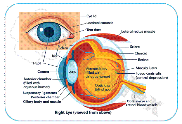
Anatomy And Physiology Review
The eye is a complex sensory organ responsible for vision. Key structures include:
- Sclera: The white outer layer that provides shape and protection.
- Cornea: A transparent, avascular layer at the front of the eye that refracts light.
- Lens: A flexible structure that focuses light on the retina.
- Iris: The colored part of the eye that controls pupil size.
- Retina: The light-sensitive layer at the back of the eye containing photoreceptor cells (rods and cones).
- Optic Nerve: Transmits visual signals from the retina to the brain.
- Aqueous Humor & Vitreous Humor: Fluids that maintain intraocular pressure (IOP) and nourish the eye.
Nursing Assessment Of The Visual System
Subjective Data
- Health History: Past and present health conditions (hypertension, diabetes), previous eye surgeries, family history of eye disorders (glaucoma, cataracts).
- Medications: Current prescription and over-the-counter medications, including eye drops.
- Functional Health Patterns:
- Changes in vision (blurriness, halos around lights, floaters, sudden vision loss).
- Pain, redness, or discharge in the eye.
- Use of corrective lenses.
- Impact of vision on daily activities (driving, reading).
Objective Data
- Visual Acuity: Assessed with a Snellen chart (distant vision) or Rosenbaum chart (near vision).
- Extraocular Muscle Function: Assessed by the six cardinal fields of gaze.
- Pupillary Response: Assessed for size, shape, equality, and reaction to light.
- Intraocular Pressure (IOP): Measured by tonometry; normal range is approximately 10−21mmHg.
- Ophthalmoscopy: Direct visualization of the retina, optic nerve, and blood vessels.
Diagnostic Procedures
Tonometry
- Purpose: Measures intraocular pressure (IOP) to screen for and monitor glaucoma.
- Procedure: A topical anesthetic may be used. The tonometer makes contact with the cornea or uses a puff of air to measure the IOP.
Slit-Lamp Microscopy
- Purpose: Provides a magnified, three-dimensional view of the eye's anterior structures.
- Procedure: The patient rests their chin on a chin rest while the examiner shines a thin beam of light into the eye. Used to detect cataracts, corneal abrasions, and other anterior segment issues.
Amsler Grid Test
- Purpose: Detects and monitors central vision changes, particularly in patients with macular degeneration.
- Procedure: The patient stares at a central dot on a grid of straight lines. Any distortion, wavy lines, or a dark spot is reported immediately.
Ophthalmoscopy
- Purpose: Direct visualization of the posterior segment.
- Procedure: The examiner uses an ophthalmoscope to shine a light into the eye and view the structures. Used to assess for retinal detachment, optic nerve damage, and diabetic retinopathy.
Common Disorders Of The Eye
Cataracts
A cataract is an opacity or cloudiness of the lens.
-
Etiology: Age-related, trauma, congenital factors, radiation exposure, or systemic diseases like diabetes.
- Clinical Manifestations: Gradual decrease in vision, blurry vision, glare, abnormal color perception, and a "milky" white appearance of the pupil in advanced stages.
- Nursing Management:
- Preoperative Care: Assess visual acuity, provide patient teaching on the surgical procedure, and administer prescribed eye drops.
- Postoperative Care: Provide discharge instructions on eye protection, administer eye drops as ordered to prevent infection and inflammation, and instruct the patient to avoid activities that increase IOP.
- Patient Teaching: Advise the patient to report any sudden vision loss, severe pain, increased redness, or discharge.
Glaucoma
Glaucoma is a group of disorders characterized by increased IOP and optic nerve damage, leading to progressive peripheral vision loss.
- Etiology: Primary open-angle glaucoma (POAG) is the most common form, caused by a decrease in fluid outflow. Primary angle-closure glaucoma (PACG) is an ocular emergency caused by sudden blockage of aqueous humor outflow.
- Clinical Manifestations:
- POAG: Develops slowly with no symptoms in early stages. Patients may lose peripheral vision slowly and are often unaware of the loss until late in the disease.
- PACG: Severe, sudden pain, nausea, vomiting, blurred vision, and halos around lights.
- Nursing Management:
- Medications: Administer topical eye drops as prescribed to decrease IOP.
- Patient Teaching: Emphasize the importance of lifelong adherence to eye drop regimens, teach proper instillation technique, and explain the need for regular follow-up visits.
Retinal Detachment
This is a separation of the retinal layers from the underlying choroid. It is a medical emergency.
- Etiology: Retinal tears, trauma, or diabetic retinopathy.
- Clinical Manifestations: Sudden onset of "floaters," light flashes, and a "curtain" or "veil" over a portion of the visual field. There is no pain.
- Nursing Management:
- Preoperative: Provide emotional support, keep the patient calm, and position the patient as ordered.
- Postoperative: Position the patient as ordered to promote reattachment, administer pain medications, and provide detailed teaching on activity restrictions.
Age-Related Macular Degeneration (AMD)
A progressive degeneration of the macula, the area of the retina responsible for central vision.
- Etiology: A complex condition linked to aging, genetics, and environmental factors.
- Clinical Manifestations: Gradual loss of central vision, blurring, and distortion of straight lines.
- Nursing Management:
- Patient Teaching: Advise patients to use the Amsler grid daily to monitor for changes in vision. Encourage a diet rich in antioxidants.
- Assistive Devices: Recommend low-vision aids and adaptive devices to improve quality of life.
Other Important Eye Disorders
Dry Eye Syndrome
-
Etiology: Decreased tear production or increased tear evaporation.
-
Clinical Manifestations: Burning, itching, foreign body sensation, and redness.
-
Nursing Management: Teach proper administration of artificial tears or lubricating drops, and advise patients to avoid environments with low humidity.
Keratitis
-
Etiology: Inflammation of the cornea, often caused by infection or trauma.
-
Clinical Manifestations: Severe pain, photophobia, tearing, and foreign body sensation.
- Nursing Management: Administer prescribed topical antibiotics or antivirals and educate on proper contact lens hygiene if applicable.
Uveitis
-
Etiology: Inflammation of the uvea, which can be caused by infection, autoimmune disorders, or trauma.
-
Clinical Manifestations: Pain, redness, light sensitivity, and blurry vision.
-
Nursing Management: Administer corticosteroids as ordered to reduce inflammation, and provide pain management.
Pharmacologic Management
β-Adrenergic Blockers
- Examples: Timolol, betaxolol.
- Action: Decrease IOP by reducing the production of aqueous humor.
- Nursing Considerations: Can cause systemic effects like bradycardia, bronchospasm, and hypotension. Monitor heart rate and blood pressure, and use with caution in patients with asthma or COPD.
Carbonic Anhydrase Inhibitors
- Examples: Dorzolamide, acetazolamide.
- Action: Decrease IOP by reducing aqueous humor production.
- Nursing Considerations: Acetazolamide can cause systemic side effects like paresthesia and electrolyte imbalances.
Prostaglandin Agonists
- Examples: Latanoprost, travoprost.
- Action: Increase the outflow of aqueous humor.
- Nursing Considerations: Can cause a harmless darkening of the iris color and increased eyelash growth.
Key Nursing Diagnoses
- Disturbed Sensory Perception (Visual) related to altered sensory reception or transmission as evidenced by a decline in visual acuity.
- Risk for Injury related to visual impairment as evidenced by difficulty ambulating and reading.
- Anxiety related to loss of vision or fear of disease progression as evidenced by verbalizing worry and restlessness.
- Deficient Knowledge related to the disease process and treatment regimen as evidenced by questions about medication and follow-up care.
Summary
- Nursing Assessment: Includes both subjective and objective data to evaluate visual function, eye health, and risk factors.
- Key Diagnostic Procedures: Tonometry for IOP, slit-lamp for anterior structures, Amsler grid for central vision, and ophthalmoscopy for the posterior segment.
- Pharmacological Management: Medications like β-blockers and carbonic anhydrase inhibitors decrease IOP, while prostaglandin agonists increase aqueous humor outflow.
- Nursing Role: Focuses on patient education, medication administration, postoperative care, and emotional support to manage symptoms and prevent disease progression
Naxlex
Videos
Login to View Video
Click here to loginTake Notes on Disorders of the Eye
This filled cannot be empty
Join Naxlex Nursing for nursing questions & guides! Sign Up Now
